
A structured overview of blood collections near the pregnancy tissues
A pregnancy hematoma is a collection of blood that forms near the pregnancy structures. It is usually detected on ultrasound and may be found after vaginal bleeding, spotting, cramping, pelvic discomfort or sometimes without symptoms at all.
For many women, the word hematoma sounds alarming. But the meaning depends on context.
A small blood collection found early in pregnancy is not the same as a large collection behind the placenta later in pregnancy. A hematoma near the membranes is not the same as bleeding that suggests placental separation. Location, size, symptoms, gestational age and change over time all matter.
This is why a pregnancy hematoma should not be understood only as “a clot” or “a bleed.” It should be understood anatomically.
Where is the blood located?
That question often matters as much as the fact that blood is present.
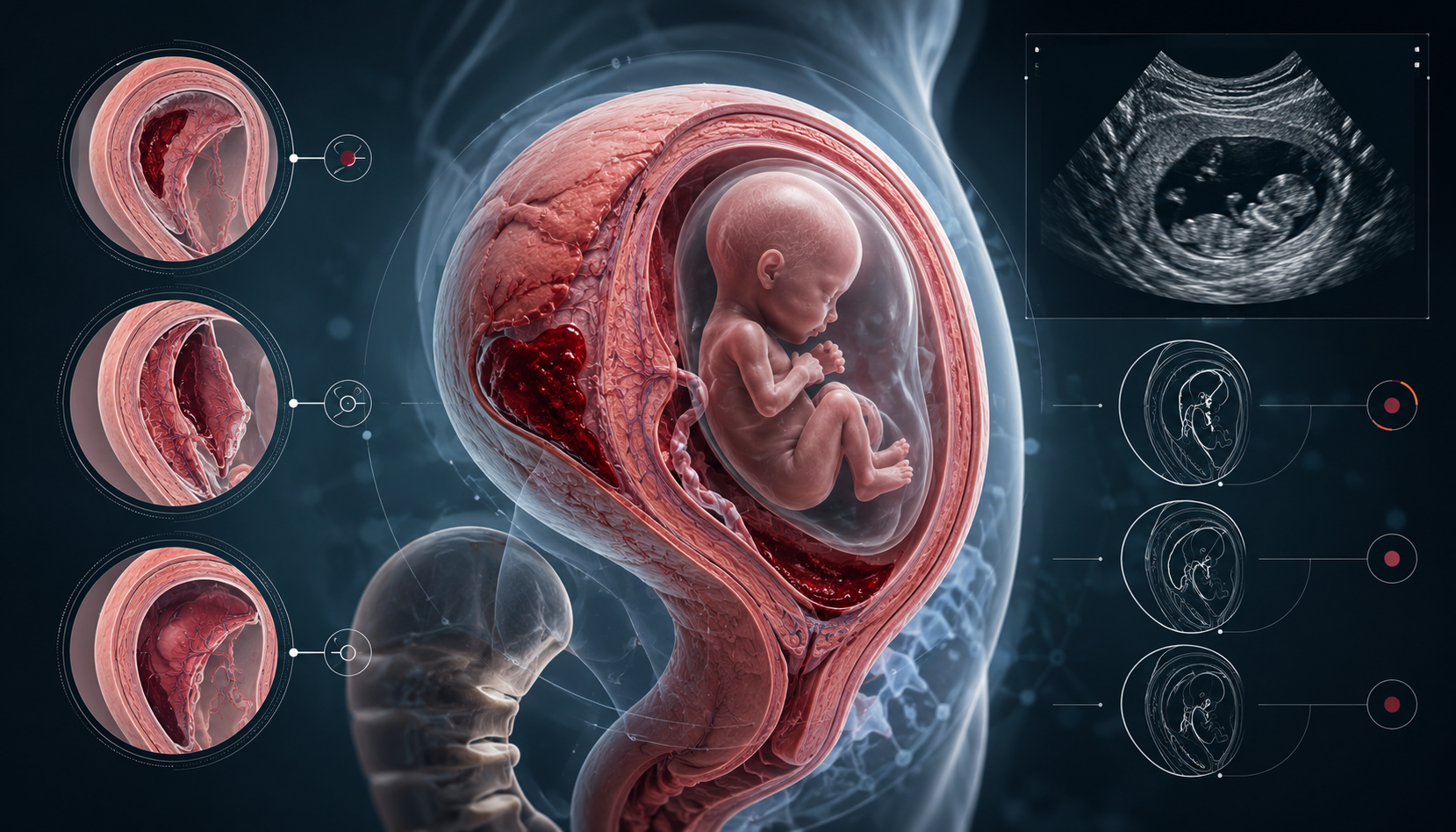
What is a pregnancy hematoma?
A hematoma is a localized collection of blood outside a blood vessel.
In pregnancy, this blood may collect between or near structures such as:
- the uterine wall
- the decidua, which is the pregnancy-adapted lining of the uterus
- the chorion and other gestational membranes
- the placenta
- the edge of the placenta
On ultrasound, a hematoma may appear as a dark or mixed-density area near the gestational sac, membranes or placenta. Its appearance can change over time as the blood ages, organizes and is gradually absorbed or drains.
Some hematomas are found because a woman has bleeding. Others are found incidentally during a routine ultrasound.
The important point is that “hematoma” describes a finding. It does not automatically describe the cause, severity or outcome.
Why location matters
Location matters because the pregnancy is made of different tissue interfaces.
Blood can collect:
- between the chorion and the uterine lining
- near the edge of the placenta
- behind the placenta
- near the membranes
- close to the cervix
- in a position that may or may not affect the placental attachment
These are not identical situations.
A small hematoma at the edge of the membranes may behave differently from a large retroplacental hematoma. A hematoma found in the first trimester may have a different meaning than a persistent or newly detected collection in the second trimester. A stable hematoma may be less concerning than one that grows or is associated with heavy bleeding, pain or uterine tenderness.
This is why ultrasound reports often describe location, size and relationship to the placenta.
Patients often focus on the word “hematoma.” Clinicians usually want to know more:
Where is it?
How large is it?
Is it changing?
Is there bleeding?
Is there pain?
Where is the placenta?
How far along is the pregnancy?
Is fetal growth and wellbeing reassuring?
Common anatomical terms
Subchorionic hematoma
A subchorionic hematoma is usually described as a blood collection between the chorion, which is part of the outer pregnancy membrane, and the uterine lining.
This is one of the most commonly discussed forms of pregnancy hematoma, especially in early pregnancy.
It may be associated with spotting or bleeding, but it can also be found without symptoms.
Many subchorionic hematomas become smaller over time, but the course varies. Some disappear quickly. Others remain visible for weeks. Some may change shape or appearance as the blood organizes.
Marginal or subplacental hematoma
A marginal hematoma is located near the edge of the placenta. It may be described as marginal, subplacental or adjacent to the placental margin depending on how it appears and how the report is written.
This location is important because it is closer to the placental attachment area than a small isolated membrane collection.
The clinical significance depends on size, symptoms, gestational age and whether there are signs of placental involvement.
Retroplacental hematoma
A retroplacental hematoma is located behind the placenta, between the placenta and the uterine wall.
This location can be more concerning because it may suggest bleeding at the placental attachment site. In some cases, retroplacental bleeding may be associated with placental abruption, although not every ultrasound finding has the same meaning.
A retroplacental hematoma should be interpreted carefully by the responsible healthcare team, especially if there is pain, uterine tenderness, contractions, heavy bleeding or fetal concerns.
Symptoms: what may be noticed
A pregnancy hematoma may cause:
- light spotting
- heavier vaginal bleeding
- passage of older brown blood
- cramping
- pelvic discomfort
- no symptoms at all
Brown discharge can sometimes represent older blood leaving the uterus. Red bleeding may suggest more recent bleeding. However, bleeding color alone cannot determine severity.
Some women have dramatic bleeding and still continue with a healthy pregnancy. Others have little bleeding but need closer follow-up because of the hematoma’s location or other risk factors.
Symptoms must be interpreted together with ultrasound findings and clinical assessment.
What ultrasound can and cannot tell us
Ultrasound is central because it can help identify where the blood collection is located and how it relates to the membranes, placenta and cervix.
Ultrasound may describe:
- hematoma size
- location
- appearance
- relationship to the placenta
- relationship to the gestational sac or membranes
- fetal heartbeat and movement
- placental position
- cervical length or appearance when relevant
- change compared with previous scans
But ultrasound does not always explain why the hematoma formed.
In many cases, the exact cause remains uncertain. Possible contributing factors may include small-vessel bleeding, partial membrane separation, placental interface changes, inflammation, trauma, coagulation factors or underlying reproductive history. In many pregnancies, no single cause is identified.
This uncertainty matters. It is one reason why hematoma information should be communicated carefully.
Size and change over time
Size matters, but size alone is not everything.
A hematoma may be described by measurements in three dimensions, by estimated volume, or by its size relative to the gestational sac or placenta. Different clinics and studies may use different methods, which can make comparisons difficult.
Change over time can be clinically important.
Questions include:
Is the hematoma shrinking?
Is it stable?
Is it growing?
Is the bleeding continuing?
Is it changing from fresh-looking blood to older organized blood?
Is fetal growth appropriate?
Is the placenta otherwise reassuring?
Some hematomas resolve quickly. Others persist for weeks or months. Persistence does not automatically mean worsening, but it does mean the finding should be interpreted in context.
Why some hematomas take time to resolve
Many patients imagine that a hematoma should drain like liquid through an open channel.
In reality, a pregnancy hematoma may sit in a narrow tissue interface rather than in a clear drainage pathway. Blood may clot, organize and then gradually break down. The body may absorb old blood products over time. Some blood may also leave through the cervix, causing spotting or brown discharge.
This process can be slow.
The exact clearance pathway may vary between cases. Some explanations are established, such as blood organization and gradual resorption. Other mechanical explanations, such as compression between closely opposed elastic pregnancy tissues, should be treated as proposed models rather than proven mechanisms for every case.
The honest answer is that hematoma resolution is partly understood, but not perfectly predictable.
Risk: avoiding both panic and false reassurance
Most information about pregnancy hematoma must be balanced.
It is not helpful to tell every woman that a hematoma is dangerous.
It is also not helpful to say that it never matters.
The risk depends on several factors:
- gestational age
- size
- location
- whether it is near or behind the placenta
- whether it is growing
- amount of bleeding
- pain or contractions
- fetal growth and wellbeing
- previous pregnancy history
- IVF or fertility history
- other maternal medical factors
Many pregnancies with hematoma continue normally. At the same time, some studies associate certain hematomas, especially larger or symptomatic ones, with increased risk of miscarriage, preterm birth, placental abruption or fetal growth restriction.
This does not mean that a hematoma predicts a bad outcome. It means that the finding should be interpreted thoughtfully.
When to seek medical care
Any vaginal bleeding during pregnancy should be discussed with a healthcare provider.
Urgent assessment is especially important if bleeding is heavy, if there is severe pain, dizziness, fainting, uterine tenderness, contractions, fever, fluid leakage, or decreased fetal movement later in pregnancy.
This article is educational and does not replace medical care. A pregnancy hematoma should be interpreted by the responsible clinical team with access to the full pregnancy history, examination findings and ultrasound report.
Established facts, uncertainty and research questions
Established:
A pregnancy hematoma is a real blood collection near pregnancy tissues, usually identified by ultrasound. Location, size, symptoms and gestational age are important for interpretation.
Uncertain:
The exact cause is often not known. The same ultrasound label may include different biological situations. Studies do not always agree on outcome risk, partly because hematomas differ in size, timing, location and patient background.
Open research questions:
Can hematoma persistence be predicted by location and tissue geometry?
Does inflammation at the decidual or placental interface increase risk in some women?
Can better ultrasound classification improve follow-up?
Can symptom tracking and biomarker trends help identify which cases need closer monitoring?
Can AI-supported interpretation help organize patterns without creating false certainty?
These are the kinds of questions Pregnancy Intelligence should explore.
Key message
A pregnancy hematoma is not just “blood.”
It is blood in a specific anatomical location, during a specific stage of pregnancy, with a specific pattern over time.
Understanding where it is, how it changes and what symptoms are present can make the finding easier to discuss, monitor and interpret.
The goal is not panic.
The goal is structured understanding.
References and Resources
- Subchorionic hematoma: Research status and pathogenesis — Review Medicine International / Spandidos Publications. Strong background source for definition, unclear pathogenesis, uncertain outcome data, and lack of uniform treatment guidelines.
- Bleeding During Pregnancy ACOG — American College of Obstetricians and Gynecologists. Patient safety source for bleeding in pregnancy and recommendation to contact an ob-gyn with bleeding.
- Subchorionic Hematoma: Causes, Symptoms & Treatment Cleveland Clinic Patient-friendly clinical overview of subchorionic hematoma, symptoms, diagnosis and general management.
- SMFM Consult Series #60: Management of Pregnancies Resulting from IVF Society for Maternal-Fetal Medicine — SMFM Useful for IVF pregnancy context, detailed ultrasound assessment, placental location, cord insertion and surveillance recommendations.