
A proposed mechanical model for slow hematoma resolution
Many women are told that a pregnancy hematoma will “resolve” or “be absorbed by the body.”
That may be true. But it can also feel incomplete.
If the hematoma is still visible after several weeks, if bleeding comes and goes, or if brown discharge continues after a scan, the simple explanation that “the body will clear it” may not answer the real question:
Why can it take so long?
The answer is probably not one single mechanism. A hematoma is blood in a specific anatomical space. How quickly it clears may depend on size, location, gestational age, tissue pressure, blood organization, drainage, resorption and the relationship between the membranes, placenta and uterine wall.
This article explains one possible mechanical model for why some pregnancy hematomas may persist.
It is important to say this clearly:
The basic process of blood clot organization and gradual resorption is established biology.
The idea that some hematomas persist because they are trapped in a compressed, narrow tissue interface is a proposed mechanical model.
It should not be presented as a proven mechanism for every pregnancy hematoma.
First: a hematoma is not a free liquid pocket
A pregnancy hematoma is often imagined as a pool of blood sitting in an open space.
But that may be misleading.
In many cases, the blood collection is located between tissues that are normally closely apposed: the uterine lining, decidua, chorionic membrane, placental edge or placental surface. These tissues are soft, elastic and under changing pressure as the uterus grows.
This means the hematoma may not behave like fluid in a cavity with an open drain.
It may behave more like blood trapped in a narrow potential space.
A potential space is not a large empty room. It is a space that opens only because something, such as blood, separates tissues that normally lie close together.
This distinction matters.
If there is no clear open channel, clearance may depend mostly on slow biological breakdown, gradual resorption and sometimes intermittent leakage through the cervix.
What is established: blood can organize and be resorbed
When bleeding occurs outside a blood vessel, the body usually responds by forming a clot.
Over time, the clot may:
- stabilize
- organize
- change ultrasound appearance
- break down into blood products
- be cleared by immune cells
- be gradually resorbed into surrounding tissues
- sometimes drain partially through the cervix
This is not instant.
Old blood can remain visible. Brown discharge may represent older blood leaving the uterus. Ultrasound appearance may change as the blood ages and organizes.
This helps explain why a hematoma may not disappear from one scan to the next.
A hematoma can be biologically “on its way out” and still remain visible.
The proposed mechanical model: compressed elastic tissues
The proposed model is this:
Some pregnancy hematomas may persist because the blood lies between elastic tissues that press toward each other and move together as the uterus grows.
Instead of forming a round cavity with an easy outlet, the hematoma may be compressed into a thin, slit-like pocket.
In that situation:
- the membranes and uterine wall may remain closely opposed
- the blood may be trapped in a narrow interface
- pressure from surrounding tissues may limit free drainage
- the clot may organize rather than flow out
- only small amounts may escape intermittently
- most clearance may depend on gradual resorption
This could help explain why some women see a pattern of slow change rather than rapid disappearance.
It could also help explain why bleeding may stop, return as spotting, turn brown, and then stop again.
However, this mechanical model needs to be handled carefully. It is a plausible anatomical interpretation, not a fully validated explanation for every case.
Why there may not be a clear drainage channel
Patients often ask a very reasonable question:
If there is blood, why does it not simply drain out?
Sometimes some blood does drain out. This may appear as red bleeding, brown discharge or intermittent spotting.
But not all hematomas have an obvious pathway to the cervix. Some may be located higher in the uterus, near membranes, near the placental edge or behind tissue planes where drainage is limited.
Even when some drainage occurs, the rest of the clot may remain in place and be cleared slowly.
A hematoma may therefore shrink through a combination of:
- partial drainage
- clot contraction
- tissue compression
- gradual breakdown
- resorption
- remodeling of the local tissue interface
This is why the amount of visible bleeding does not always match the size of the hematoma.
A woman can have a visible hematoma with little bleeding.
Another woman can have dramatic bleeding and later show a smaller collection.
The relationship is not always intuitive.
Why size can change slowly
The size of a hematoma may change in several ways.
It may become smaller because the blood is being absorbed. It may change shape because the uterus is growing. It may look different because the blood is becoming older and more organized. It may appear larger if there has been new bleeding or if the imaging plane captures it differently.
This is why ultrasound follow-up is not always a simple linear story.
A hematoma can:
- shrink
- remain stable
- change shape
- look more organized
- show mixed echogenicity
- appear different depending on measurement technique
- occasionally increase if new bleeding occurs
This can be frustrating for patients because they want a clear answer: better or worse?
Sometimes the answer is more nuanced.
The trend matters, but so does the clinical context.
Why weeks or months can be realistic
A pregnancy hematoma may take time because several slow processes may be happening at once.
The blood is not only “leaving.” It may first need to clot, stabilize, organize, break down and be resorbed.
If the hematoma sits in a compressed tissue plane, mechanical clearance may be limited. If there is no clear drainage route, the body may need to clear most of it gradually.
This means that weeks or even months of visibility on ultrasound can be biologically plausible.
Persistence does not automatically mean danger. But persistence should be interpreted in relation to:
- gestational age
- location
- size
- symptoms
- placental relationship
- fetal growth
- whether the hematoma is shrinking, stable or growing
- whether there is ongoing fresh bleeding
- whether there is pain, contractions or other warning signs
A stable, shrinking or organizing hematoma is different from one associated with worsening symptoms or new bleeding.
Why brown bleeding can happen later
Brown bleeding or discharge often worries patients because it can appear after a period of no bleeding.
One possible explanation is that older blood is leaving slowly.
When blood has been present for some time, it changes color as it breaks down. Brown discharge may represent older blood rather than active fresh bleeding.
However, color alone cannot prove whether bleeding is old or new. Any bleeding pattern should be interpreted with the full clinical picture.
The useful point is this:
Delayed brown discharge can be consistent with slow hematoma clearance.
It does not automatically mean something new has happened. But it also should not be ignored if it is heavy, painful, recurrent or associated with other symptoms.
Why patients need better explanations
Many women with pregnancy hematoma are told only:
“Rest.”
“Wait.”
“It will be absorbed.”
“Come back if it gets worse.”
Sometimes that is medically appropriate, but it may not be enough intellectually.
A patient who has read her ultrasound report, tracked bleeding patterns and followed hematoma measurements over time often needs a more structured explanation.
She wants to understand:
Why is it still there?
Why does old blood come out later?
Why does it change shape?
Why can it shrink slowly?
Why does one scan look different from another?
Why do doctors not always know the cause?
These are not childish questions.
They are serious questions about anatomy, biology and uncertainty.
A better explanation can reduce fear without giving false reassurance.
What remains uncertain
The exact clearance pathway for each hematoma is often not known.
Research describes associations between hematoma, bleeding and pregnancy outcomes, but the literature is not completely consistent. Studies differ in how they define hematoma, when scans are performed, how size is measured, whether symptoms are present, and what patient groups are included.
This means it is difficult to make simple statements that apply to every case.
Important uncertainties include:
- why some hematomas persist longer than others
- whether tissue geometry predicts resolution time
- how inflammation affects local bleeding and healing
- whether different hematoma locations have different clearance patterns
- how ultrasound appearance corresponds to biological healing
- whether symptom patterns can help distinguish old drainage from new bleeding
- whether AI-supported trend analysis could improve follow-up
This is why the mechanical model should be presented as a research idea, not as established clinical fact.
Practical interpretation
For patients, the most useful interpretation is often this:
A hematoma can take time to clear because blood may be trapped in a tissue interface, not sitting in an open space. The body may need to break down and resorb the clot gradually. Some blood may drain intermittently, but not every hematoma has a clear drainage pathway.
This explanation does not replace ultrasound follow-up or clinical care.
It simply gives a more realistic model of what may be happening.
When to seek medical care
Any bleeding during pregnancy should be discussed with a healthcare provider.
Urgent assessment is especially important if there is:
- heavy bleeding
- severe pain
- dizziness or fainting
- contractions
- fluid leakage
- fever
- uterine tenderness
- reduced fetal movement later in pregnancy
- a sudden change in symptoms
This article is educational and does not replace medical evaluation.
Key message
A pregnancy hematoma may take weeks or months to clear not only because the body must break down blood, but also because the blood may lie in a narrow tissue interface with limited drainage.
The biological process of clot organization and resorption is established.
The mechanical model of a compressed, slowly clearing hematoma pocket is a proposed explanation that may help patients and researchers think more clearly about persistence.
It is not proven for every case.
The goal is structured understanding, not false certainty.
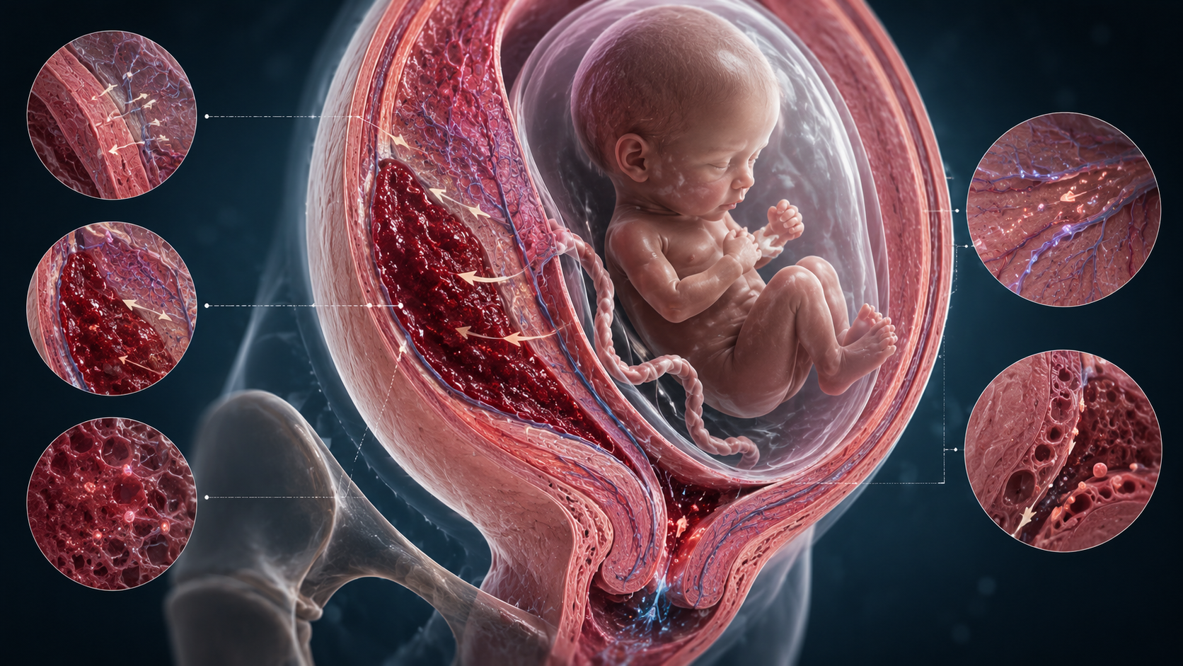
Figure: Proposed clearance pathways for a pregnancy hematoma.
The main image shows a cross-section of the uterus with the fetus inside the amniotic sac. The hematoma is visible as a dark red blood collection behind the placenta, close to the uterine wall. The surrounding tissue space is narrow rather than open, which may help explain why some hematomas persist for weeks or months.
The small images illustrate possible contributors to slow clearance: the upper left image shows gradual resorption into surrounding tissue; the middle left image shows clot organization and breakdown; the lower left image shows limited drainage toward the cervix; the upper right image shows the compressed tissue interface around the hematoma; and the lower right image shows microscopic cleanup through local tissue and vascular pathways.
This figure combines established biology, such as clot breakdown and resorption, with a proposed mechanical model. The exact clearance pathway may vary between pregnancies and is not proven for every hematoma.
References and Resources
- Subchorionic hematoma: Research status and pathogenesis Medicine International / Spandidos Publications Background on SCH pathogenesis, uncertainty, conflicting outcome data and treatment limitations. The review notes that SCH has been associated with adverse outcomes in some studies, while other studies do not find increased risk.
- Subchorionic Hematoma: Causes, Symptoms & Treatment Cleveland Clinic Patient-facing clinical overview explaining that SCH can cause vaginal bleeding and is a blood collection between pregnancy tissues and the uterine wall.
- Bleeding During Pregnancy ACOG — American College of Obstetricians and Gynecologists Safety source supporting the recommendation that bleeding during pregnancy should be discussed with an obstetric provider.
- Subchorionic hematoma and risk of preterm delivery: a systematic review and meta-analysis American Journal of Obstetrics & Gynecology MFM Research context for the mixed evidence base and outcome uncertainty in SCH studies.