
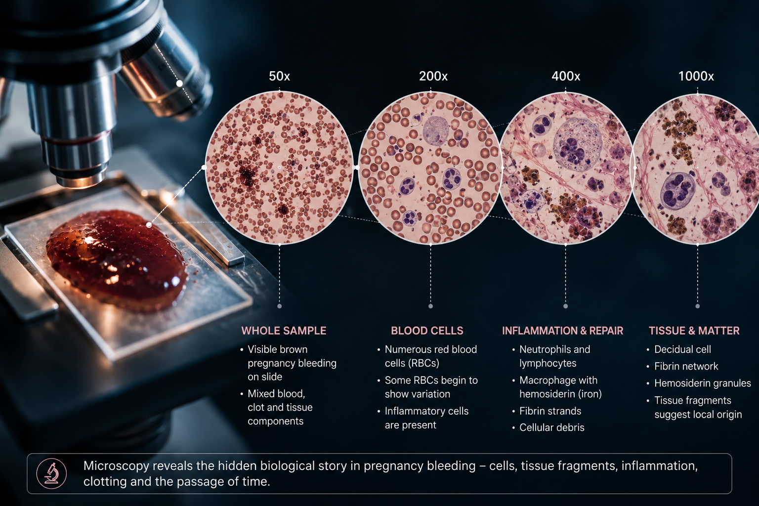
What would we see if we simply put the sample under a microscope?
A woman presents with vaginal bleeding during pregnancy.
The blood is visible.
Its color can be described.
Its quantity can be estimated.
Clots may be present.
Tissue fragments may be visible.
But what happens when we move beyond the naked eye?
What if we place a small drop of the bleeding on a glass slide and look at it under a microscope?
The answer seems simple.
We would see blood.
But that answer may be incomplete.
A pregnancy bleeding sample is often more than blood. It may contain inflammatory cells, tissue fragments, cervical cells, decidual material, fibrin, microorganisms and evidence of biological processes that occurred before the bleeding became visible.
In a forensic framework, microscopy becomes the second layer of evidence.
The question is no longer:
"Is there bleeding?"
The question becomes:
"What biological story is hidden inside the bleeding?"
Why Microscopy Matters
Every biological event leaves traces.
Inflammation leaves cells.
Tissue injury leaves fragments.
Blood aging leaves degradation products.
Healing leaves organization.
Infection leaves microorganisms and immune responses.
A microscope allows us to see some of these traces directly.
Unlike chemistry or DNA analysis, microscopy has a unique advantage:
It is visual.
Instead of measuring molecules indirectly, we can observe structures themselves.
That makes microscopy one of the oldest and most intuitive forensic tools in medicine.
The First Thing We See: Red Blood Cells
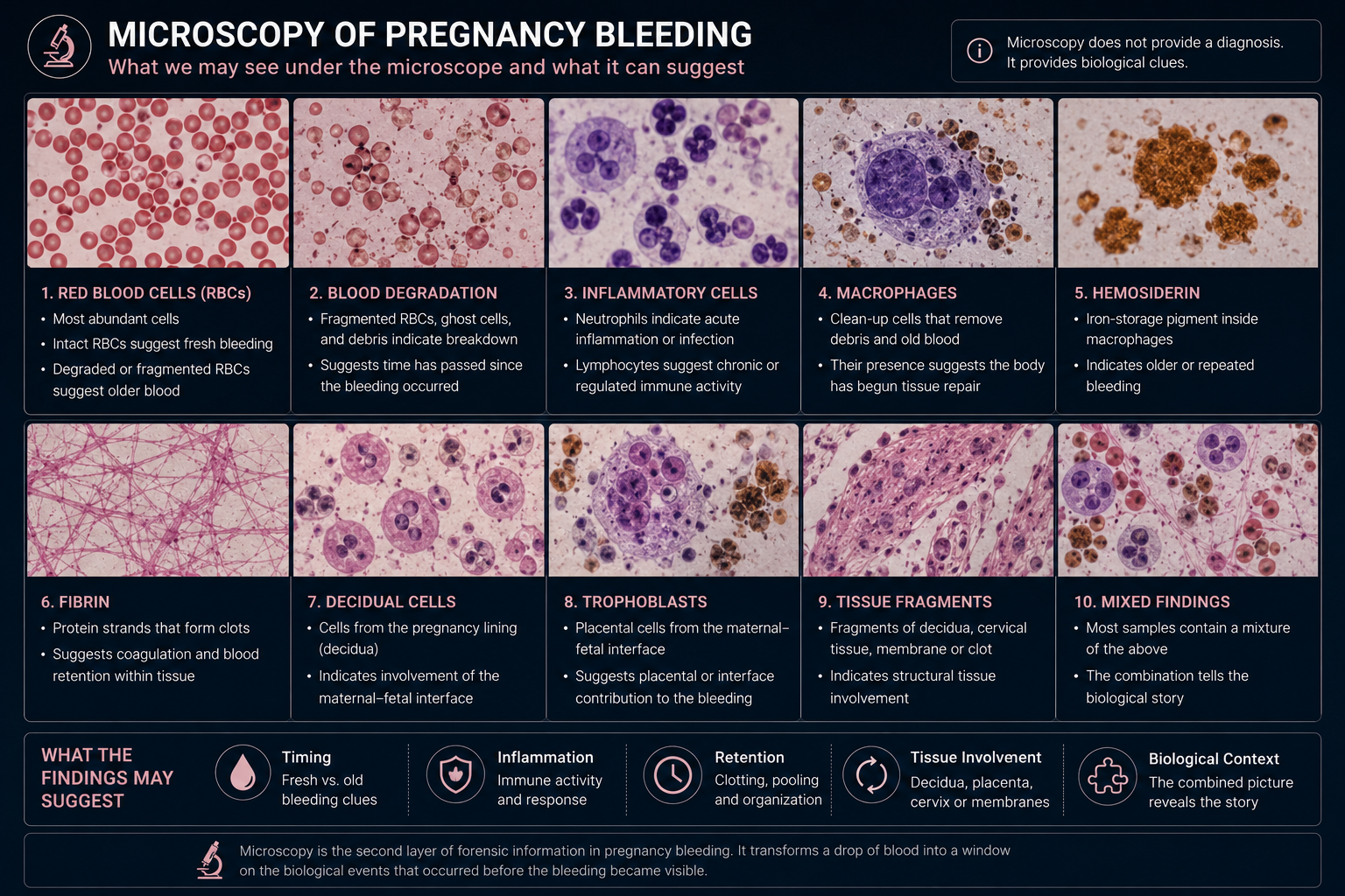
The most abundant cells in most bleeding samples are red blood cells.
Under the microscope they appear as small circular discs without nuclei.
Their primary role is oxygen transport.
At first glance they may seem uninteresting.
But their appearance can provide clues.
Fresh Bleeding
Fresh blood typically contains large numbers of intact erythrocytes.
The cells maintain their normal shape.
The sample may appear relatively clean.
Possible interpretation:
- recent bleeding
- active bleeding
- limited degradation
Older Bleeding
As blood ages, red blood cells begin to break down.
Possible findings:
- fragmented erythrocytes
- distorted shapes
- cellular ghosts
- loss of membrane integrity
- pigment accumulation
Possible interpretation:
- retained blood
- hematoma drainage
- previous bleeding event
- prolonged exposure to tissue environment
This means the red blood cells themselves may provide information about timing.
Blood Degradation: A Biological Clock
Blood does not remain unchanged.
Once outside the circulation, a sequence of degradation begins.
Hemoglobin changes.
Membranes deteriorate.
Cells rupture.
Pigments accumulate.
This creates a microscopic spectrum between fresh blood and old blood.
Possible findings include:
- intact erythrocytes
- partially degraded erythrocytes
- ghost cells
- cellular debris
- pigment deposits
A sample dominated by degraded cells may suggest a very different biological process than a sample dominated by fresh cells.
For example:
A fresh cervical bleed may look very different from a draining decidual hematoma that has existed for days or weeks.
White Blood Cells: Evidence of Inflammation
One of the most important findings in microscopy is the presence of inflammatory cells.
The body sends immune cells to sites of injury, infection and tissue remodeling.
These cells may become visible in the bleeding sample.
Neutrophils
Neutrophils are often the first responders.
Increased numbers may suggest:
- acute inflammation
- infection
- tissue injury
- active immune response
Lymphocytes
Lymphocytes may indicate more organized immune activity.
Possible interpretation:
- chronic inflammation
- immune regulation
- tissue adaptation
Eosinophils
Less common but potentially relevant in specific inflammatory environments.
Microscopy cannot always determine the exact cause.
But it can show that inflammation is present.
That alone changes the biological interpretation.
Macrophages: The Cleanup Crew
Macrophages are among the most interesting cells in forensic microscopy.
Their role is cleanup.
They remove:
- dead cells
- damaged tissue
- blood products
- debris
When bleeding has existed for some time, macrophages often begin to appear.
Possible interpretation:
- previous bleeding event
- active tissue repair
- hematoma organization
- ongoing cleanup process
In many ways, macrophages act as evidence that time has passed.
Their presence may suggest that the body has already begun responding to the bleeding.
Hemosiderin: Evidence of Old Blood
One of the classic markers of previous bleeding is hemosiderin.
Hemosiderin is an iron-storage pigment that forms during the breakdown of hemoglobin.
Macrophages may accumulate hemosiderin after ingesting red blood cells.
Under appropriate staining methods, these cells can become visible.
Possible interpretation:
- older bleeding
- repeated bleeding
- chronic blood degradation
- resolving hematoma
In forensic pathology, hemosiderin is often viewed as evidence that blood has been present long enough for breakdown and cleanup to begin.
For pregnancy bleeding, this could help distinguish fresh events from older organized processes.
Fibrin: The Architecture of Clotting
When blood coagulates, fibrin is formed.
Fibrin acts as a structural scaffold.
Under the microscope it may appear as:
- strands
- networks
- mesh-like structures
Possible interpretation:
- clot formation
- coagulation activity
- blood retention
- hematoma organization
A sample rich in fibrin may indicate that blood has not simply flowed out immediately.
It may have spent time organizing within tissue.
Decidual Cells: Evidence from the Pregnancy Lining
The decidua is the specialized uterine lining present during pregnancy.
If decidual tissue contributes to the bleeding, decidual cells may become visible.
Possible interpretation:
- decidual involvement
- bleeding near the maternal-fetal interface
- local tissue disruption
- decidual inflammation
This is particularly interesting because decidual biology sits at the center of implantation, placentation and immune regulation.
Finding decidual material transforms the sample from a simple blood specimen into a tissue specimen.
Trophoblasts: Clues from the Placental Interface
Trophoblasts are placental cells involved in the connection between mother and fetus.
Their presence in a bleeding sample could suggest involvement of the placental interface.
Possible interpretation:
- placental contribution
- maternal-fetal interface activity
- tissue remodeling
- local disruption near placental structures
Identification of trophoblasts can be challenging and may require additional techniques.
Nevertheless, they represent one of the most interesting potential findings because they connect directly to placental biology.
Tissue Fragments: Structural Evidence
Microscopy may reveal fragments larger than individual cells.
These fragments may contain:
- decidual tissue
- cervical tissue
- membrane material
- fibrin
- cellular aggregates
Possible interpretation:
- structural tissue involvement
- active remodeling
- tissue disruption
- healing process
Tissue fragments often raise more questions than answers.
But they can point investigators toward specific biological regions.
A Fresh Bleed Versus an Old Hematoma
Imagine two patients.
Sample A
Microscopy shows:
- abundant intact erythrocytes
- minimal debris
- few inflammatory cells
- little fibrin
Possible interpretation:
Recent bleeding.
Sample B
Microscopy shows:
- degraded erythrocytes
- macrophages
- hemosiderin
- fibrin
- cellular debris
Possible interpretation:
Older retained blood, hematoma organization or prolonged degradation.
Both patients may report vaginal bleeding.
Yet microscopically they may represent very different biological events.
The Microscopy Framework
A structured microscopy assessment could ask:
Red Blood Cells
- Intact or degraded?
- Fresh or aged appearance?
Inflammatory Cells
- Few or many?
- Acute or chronic pattern?
Macrophages
- Present or absent?
- Evidence of cleanup?
Hemosiderin
- Evidence of older bleeding?
Fibrin
- Evidence of clot organization?
Decidual Material
- Evidence of pregnancy lining involvement?
Trophoblast Material
- Evidence of placental interface involvement?
Tissue Fragments
- Evidence of structural tissue disruption?
This transforms microscopy from a descriptive exercise into a biological reconstruction tool.
What Microscopy Cannot Tell Us
Microscopy is powerful but limited.
It cannot reliably determine:
- exact timing
- fetal condition
- precise molecular pathways
- genetic risk
- complete tissue origin
For those questions we need chemistry, proteomics, DNA analysis and other methods.
But microscopy provides something unique.
It allows us to see biology directly.
The Central Question
A microscope does not merely show blood.
It shows cells.
It shows damage.
It shows repair.
It shows inflammation.
It shows the passage of time.
The central question is therefore not:
"Is there blood?"
It is:
"What happened to the blood before it reached the microscope?"
That is where forensic interpretation begins.
Conclusion: The Second Layer of Evidence
Visual inspection asks:
What does the bleeding look like?
Microscopy asks:
What is actually inside the bleeding?
Red blood cells, inflammatory cells, macrophages, hemosiderin, fibrin, decidual tissue and trophoblast-derived material may all provide clues.
None of these findings alone provide a diagnosis.
But together they begin transforming a drop of blood into a biological narrative.
A bleeding sample may not simply be evidence that bleeding occurred.
It may be evidence of how, where and when it occurred.
And that makes microscopy the second layer in the growing framework of Pregnancy Bleeding Intelligence.
References and Resource
1. The Role of Decidual Cells in Uterine Hemostasis, Menstruation, Inflammation and Abnormal Uterine Bleeding Human Reproduction Update / Oxford Academic
A useful review on decidual biology, hemostasis and bleeding-related mechanisms in reproductive tissues. It supports the idea that decidual tissue is not passive, but actively involved in bleeding control, inflammation and tissue remodeling.
2. Placenta – Maternal Decidua UNSW Embryology
A clear educational resource explaining the maternal decidua, trophoblast invasion and the maternal-fetal interface. Useful background for understanding why decidual cells or trophoblast-related material may be relevant in pregnancy bleeding microscopy.
3. Fibrin Architecture in Clots: A Quantitative Polarized Light Microscopy Study PLOS ONE / PubMed Central
Explains how fibrin forms an important structural component of clots and how microscopy can be used to study clot architecture. Relevant for interpreting fibrin strands and clot organization in older or retained bleeding.
4. Local Immune Recognition of Trophoblast in Early Human Pregnancy Nature Reviews Immunology
A high-quality review of trophoblast interaction with the maternal immune system at the decidual interface. Useful for the article’s discussion of trophoblasts, immune cells and maternal-fetal interface biology.
5. Hemosiderin-Laden Macrophages and Prior Hemorrhage University of Utah WebPath
Educational pathology reference showing how macrophages can contain brown hemosiderin pigment after red blood cell breakdown. Useful for explaining why hemosiderin-laden macrophages suggest older or repeated bleeding.