
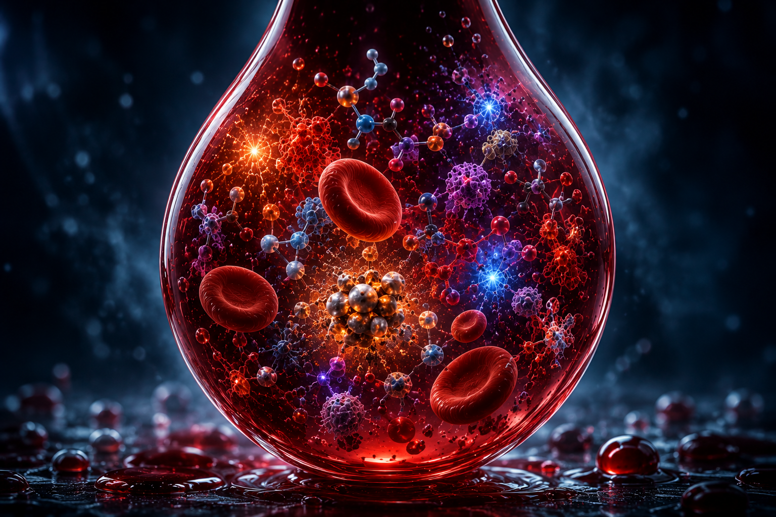
What molecules are dissolved in the sample?
A woman presents with vaginal bleeding during pregnancy.
The blood is visible.
Under the microscope we may observe cells, fibrin, inflammatory activity and tissue fragments.
Microbiology may reveal bacterial communities and microbial patterns.
But another question remains:
What chemistry is hidden inside the sample?
Blood is not simply a collection of cells.
It is also a solution.
Dissolved within that solution are thousands of molecules.
Some are involved in inflammation.
Some are involved in coagulation.
Some reflect oxidative stress.
Some are released from damaged tissue.
Some may originate from the placenta, decidua, membranes or immune system.
Together, these molecules form a chemical fingerprint.
And that fingerprint may contain clues about the biological processes occurring before the bleeding became visible.
This article explores the chemistry hidden within pregnancy bleeding.
A Bleeding Sample Is a Chemical Environment
When we see bleeding, our attention naturally focuses on red blood cells.
But cells are only part of the story.
Surrounding those cells is fluid.
That fluid contains:
- ions
- proteins
- metabolites
- inflammatory mediators
- hormones
- lipids
- enzymes
- degradation products
- signaling molecules
Every biological process leaves chemical traces.
Inflammation changes chemistry.
Infection changes chemistry.
Tissue injury changes chemistry.
Healing changes chemistry.
The question becomes:
Can we learn something about the event by studying those chemical traces?
The First Chemical Question: pH
One of the simplest measurements is pH.
pH reflects acidity and alkalinity.
The vaginal environment is normally acidic, largely because of Lactobacillus activity.
When the local ecosystem changes, pH may change as well.
Possible influences include:
- microbiome composition
- infection
- inflammation
- fluid mixing
- tissue breakdown
pH alone cannot explain bleeding.
But it provides context.
In forensic terms, it helps describe the environment in which the sample existed.
Iron Metabolism: Evidence of Blood Breakdown
Blood contains enormous amounts of iron.
Most of it is locked inside hemoglobin.
When red blood cells break down:
- hemoglobin is released
- iron metabolism changes
- degradation products accumulate
Potential findings include:
- free hemoglobin
- heme
- ferritin
- hemosiderin-related products
- iron-binding proteins
These molecules may provide clues about:
- fresh bleeding
- old bleeding
- retained blood
- hematoma degradation
- oxidative stress
In a resolving hematoma, iron chemistry may become particularly important.
The breakdown of blood is itself a chemical process.
Oxidative Stress: The Chemistry of Damage
One of the most intriguing areas of reproductive biology is oxidative stress.
Oxidative stress occurs when reactive oxygen species exceed the body's ability to neutralize them.
This can affect:
- cell membranes
- proteins
- DNA
- mitochondria
- extracellular matrix
Oxidative stress has been implicated in:
- placental dysfunction
- inflammation
- tissue injury
- abnormal implantation
- membrane weakening
A bleeding sample may contain markers reflecting these processes.
Possible examples include:
- lipid peroxidation products
- oxidized proteins
- reactive oxygen species byproducts
- antioxidant depletion signals
In simple terms:
Chemistry may reveal whether the surrounding tissues were experiencing biological stress before the bleeding occurred.
Lactate: A Marker of Metabolic Activity
Lactate is often associated with exercise.
But lactate is a universal biological molecule.
It increases when tissues shift toward anaerobic metabolism.
Possible causes include:
- inflammation
- hypoxia
- tissue stress
- immune activation
- microbial metabolism
A bleeding sample with elevated lactate may reflect a tissue environment under strain.
The challenge is interpretation.
Lactate is rarely specific.
But in combination with other findings, it may help build a broader picture.
Prostaglandins: Molecules That Change Tissues
Prostaglandins are among the most important signaling molecules in reproductive biology.
They influence:
- inflammation
- blood vessels
- uterine activity
- cervical remodeling
- membrane biology
Researchers have long linked prostaglandins to labor pathways.
They also participate in many inflammatory processes.
Potential significance:
- tissue activation
- local inflammation
- remodeling processes
- uterine signaling
A bleeding sample may therefore contain clues about biological activity occurring around the maternal-fetal interface.
Cytokines: The Language of Inflammation
If cells communicate, cytokines are part of the language.
Cytokines are signaling proteins used by immune cells and tissues.
They help coordinate responses to:
- infection
- injury
- tissue remodeling
- stress
Examples include:
- IL-1
- IL-6
- IL-8
- TNF-alpha
- IL-10
Different cytokine patterns may reflect different biological environments.
Some patterns are more inflammatory.
Some are more regulatory.
Some suggest active tissue injury.
Some suggest healing.
In a forensic framework, cytokines may help answer:
Was inflammation present before the bleeding became visible?
Chemokines: The Recruitment Signals
Chemokines are closely related to cytokines.
Their primary role is attracting cells.
They tell immune cells where to go.
When tissue becomes inflamed, chemokines often increase.
This leads to recruitment of:
- neutrophils
- macrophages
- lymphocytes
- other immune cells
Microscopy may show those cells.
Chemistry may reveal the signals that attracted them.
This is an important concept.
Microscopy shows the outcome.
Chemistry may reveal the process.
Lipid Mediators: The Forgotten Signals
Many inflammatory signals are derived from fats.
These molecules are often called lipid mediators.
Examples include:
- prostaglandins
- leukotrienes
- specialized pro-resolving mediators
These molecules influence:
- inflammation
- vascular permeability
- immune activity
- tissue repair
- resolution of injury
In recent years, lipid mediators have become a major area of research.
They may help explain why some inflammatory responses resolve successfully while others become chronic.
The Chemical Signature of a Hematoma
Imagine blood becoming trapped within tissue.
The blood does not remain unchanged.
Cells break down.
Iron is released.
Inflammatory pathways activate.
Oxidative stress develops.
Immune cells arrive.
Tissue remodeling begins.
The result is not merely old blood.
It is a changing chemical environment.
A hematoma is therefore not only an anatomical structure.
It is also a biochemical ecosystem.
This perspective may help explain why hematomas can evolve over time rather than behaving as static collections of blood.
Fresh Bleeding Versus Old Bleeding
Chemically, these may look very different.
Fresh Bleeding
Possible characteristics:
- intact hemoglobin
- limited degradation
- lower levels of breakdown products
- less evidence of cleanup
Older Bleeding
Possible characteristics:
- iron release
- heme breakdown
- oxidative stress
- inflammatory signaling
- tissue remodeling markers
Two samples may appear visually similar.
Chemically they may represent entirely different stages of a biological process.
The Forensic Chemistry Framework
A structured analysis could ask:
Environmental Chemistry
- pH
- ionic balance
- fluid composition
Blood Degradation
- hemoglobin
- heme
- ferritin
- iron-related molecules
Oxidative Stress
- oxidation products
- antioxidant balance
- lipid peroxidation markers
Metabolic Activity
- lactate
- energy metabolites
- tissue stress markers
Inflammation
- cytokines
- chemokines
- inflammatory proteins
Lipid Signaling
- prostaglandins
- leukotrienes
- specialized mediators
Tissue Remodeling
- extracellular matrix breakdown products
- remodeling signals
- repair-associated molecules
Each layer adds information.
Together they form a chemical portrait.
What Chemistry Cannot Tell Us
Chemistry is powerful.
But it is not a diagnosis.
A chemical signal may be:
- specific
- nonspecific
- protective
- harmful
- adaptive
- secondary
No molecule should be interpreted in isolation.
Chemistry becomes most useful when combined with:
- visual appearance
- microscopy
- microbiology
- proteomics
- clinical context
The goal is not to find one magic biomarker.
The goal is to understand patterns.
The Central Question
The traditional question is:
"What cells are present?"
The chemical question is different:
"What processes are occurring?"
Cells are evidence.
Chemistry is activity.
Chemistry may reveal:
- inflammation
- oxidative stress
- tissue breakdown
- repair
- immune signaling
- metabolic adaptation
These processes often begin before structural changes become obvious.
That is why chemistry is so interesting.
Conclusion: The Fourth Layer of Evidence
Visual forensics asks:
What does the bleeding look like?
Microscopy asks:
What cells and tissues are present?
Microbiology asks:
What microbial ecosystem surrounds the event?
Chemistry asks:
What biological processes are occurring?
A pregnancy bleeding sample may contain a hidden chemical archive.
Within that archive are signals from:
- iron metabolism
- oxidative stress
- inflammation
- tissue remodeling
- immune communication
- metabolic activity
Those signals may help explain not only that bleeding occurred, but why the biological environment allowed it to occur.
In the framework of Pregnancy Bleeding Intelligence, chemistry becomes the fourth layer of evidence.
Not because molecules provide all the answers.
But because biological processes leave chemical traces.
And chemistry is often where those traces become visible.
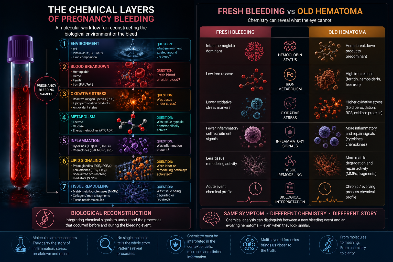
Figure 1. The Microbiome Hidden in Pregnancy Bleeding.
Conceptual framework illustrating how pregnancy bleeding may contain microbial information reflecting the vaginal and cervical microbiome. Potential findings include Lactobacillus-dominant communities, bacterial vaginosis-associated organisms, Ureaplasma species, inflammatory microbiome patterns and microbial signatures linked to membrane weakening, ascending infection and preterm birth pathways. This figure is a simulated educational illustration and is not derived from a real patient sample.
References and Resource
1. Quantification of the Size of Subchorionic Hematoma Causing Pregnancy-Related Complications: A Retrospective Cohort Study Journal of Medical Ultrasonics, published 27 August 2024.
A retrospective study examining how subchorionic hematoma size relates to pregnancy-related complications such as miscarriage, preterm delivery, preterm PROM, fetal growth restriction and placental abruption.
2. The Role of Decidual Cells in Uterine Hemostasis, Menstruation, Inflammation, Adverse Pregnancy Outcomes and Abnormal Uterine Bleeding Human Reproduction Update, published 21 June 2016.
A review of how decidual cells regulate hemostasis, inflammation, thrombin signaling, matrix remodeling and bleeding in both pregnancy and menstruation.
3. Repeated Measures of Cervicovaginal Cytokines During Healthy Pregnancy American Journal of Perinatology, published 2019.
A study measuring 20 cervicovaginal cytokines repeatedly during pregnancy, showing that local reproductive tract fluids can contain measurable inflammatory signaling patterns.
4. Decidual Inflammation Drives Chemokine-Mediated Immune Infiltration Contributing to Term Labor The Journal of Immunology, published 15 October 2021.
A study showing that decidual tissue can produce cytokines and chemokines that activate immune-cell recruitment into uterine tissues, supporting the idea that local chemistry reflects active tissue biology.
5. Involvement of Human Decidual Cell-Expressed Tissue Factor in Uterine Hemostasis and Abruption Thrombosis Research, published 2009.
A paper on how decidual tissue factor supports hemostasis during pregnancy and how disturbances in this system may relate to decidual hemorrhage and placental abruption biology.