
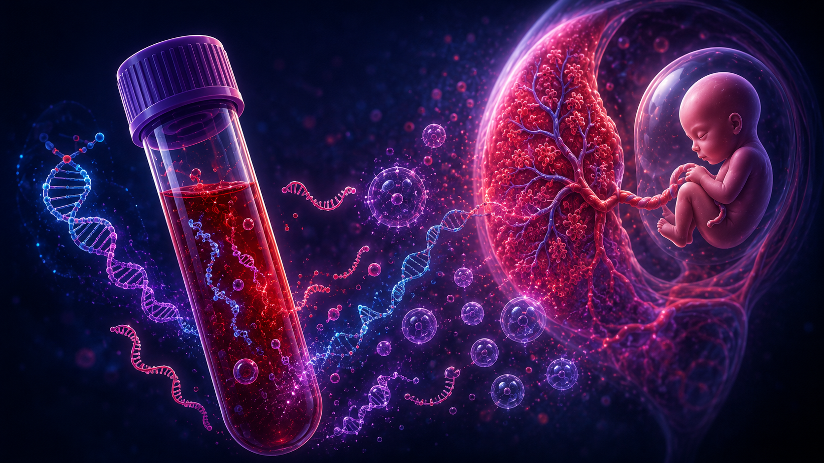
Can we identify where the bleeding originated?
Pregnancy bleeding can be seen with the eye.
It can be examined under a microscope.
It can be studied for microbes.
It can be analyzed chemically.
It can be profiled for proteins.
But DNA and RNA add another layer.
They may allow us to ask a more precise question:
Can we identify which tissue the bleeding came from?
This is the central idea behind tissue-of-origin mapping.
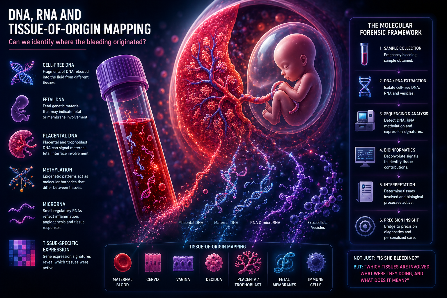
A bleeding sample may contain genetic and molecular traces from several biological sources:
- maternal blood
- cervix
- vagina
- decidua
- placenta
- fetal membranes
- fetal cells
- immune cells
- microbial organisms
Some of these sources may look similar under the microscope.
Some may share proteins.
But at the DNA, RNA and epigenetic level, tissues may carry distinct molecular fingerprints.
That makes molecular mapping one of the most important future layers in Pregnancy Bleeding Intelligence.
From Blood Sample to Molecular Evidence
A pregnancy bleeding sample is not simply red blood cells.
It may contain:
- intact maternal cells
- fetal cells
- placental cells
- decidual cells
- cervical cells
- cell-free DNA
- cell-free RNA
- microRNAs
- methylation patterns
- tissue-specific gene-expression signals
Some material may come from intact cells.
Some may come from damaged cells.
Some may be released into the fluid as fragments.
This matters because bleeding is often a mixed sample.
The sample may not have one origin.
It may contain several biological contributors.
The forensic question becomes:
Can we deconvolute that mixture?
What Is Tissue-of-Origin Mapping?
Tissue-of-origin mapping means trying to determine which tissues contributed biological material to a sample.
In cancer research, this concept is already important.
A blood sample may contain cell-free DNA from a tumor. Researchers then try to determine where that DNA came from.
The same logic can be applied conceptually to pregnancy bleeding.
A bleeding sample could contain DNA or RNA from:
- maternal blood cells
- placental trophoblasts
- decidual tissue
- fetal membranes
- cervix
- vagina
If each tissue has a different molecular signature, then a mixed sample may be partly reconstructed.
The goal is not just to say:
“There is blood.”
The goal is to ask:
Which tissues left molecular evidence in the sample?
Cell-Free DNA: Fragments Floating in Fluid
Cell-free DNA is DNA that is not contained inside intact cells.
It exists as fragments in biological fluids.
During pregnancy, maternal blood contains cell-free DNA from both the mother and the placenta.
This is the scientific basis of non-invasive prenatal testing.
But cell-free DNA is not only useful for fetal chromosome testing.
It also represents a broader biological principle:
Tissues shed DNA fragments.
If a tissue is stressed, inflamed, remodeling or damaged, it may release more DNA.
In pregnancy bleeding, cell-free DNA could potentially provide clues about:
- maternal contribution
- fetal or placental contribution
- tissue injury
- cell death
- inflammation
- interface disruption
The sample becomes molecular evidence.
Fetal DNA
Fetal DNA is usually discussed in the context of prenatal screening.
But in a bleeding sample, fetal DNA may have another possible meaning.
Its amount and context may help indicate whether fetal or placental compartments contributed material to the sample.
Important questions include:
- Is fetal DNA present?
- Is the fetal fraction unusually high?
- Is fetal DNA mixed with placental markers?
- Does the pattern suggest fetal membrane involvement?
- Does it reflect normal pregnancy shedding or tissue disruption?
Fetal DNA alone would not prove the source of bleeding.
But in combination with other layers, it could add important context.
Placental DNA
Much of what is often called fetal cell-free DNA in maternal blood is actually placental in origin.
The placenta releases DNA fragments into maternal circulation.
This is important because the placenta has distinct molecular features.
Placental DNA may carry signatures that differ from maternal blood cells or cervical tissue.
In pregnancy bleeding, placental DNA could be especially relevant if the bleeding involves:
- trophoblast tissue
- placental edge
- maternal-fetal interface
- decidual-placental separation
- hematoma formation
- placental stress
The key idea is not that placental DNA proves a specific diagnosis.
The key idea is that placental molecular material may help indicate whether the placenta or trophoblast interface is part of the bleeding event.
Methylation: The Molecular Barcode
DNA sequence tells us the genetic code.
But tissues with the same DNA sequence can behave very differently.
That is partly because of epigenetics.
One major epigenetic mechanism is DNA methylation.
Methylation patterns help regulate which genes are active or silent.
Different tissues have different methylation profiles.
This makes methylation powerful for tissue-of-origin mapping.
A blood cell, placental cell, cervical cell and decidual cell may share the same genome, but their methylation patterns can differ.
This means a bleeding sample might theoretically be analyzed for methylation signatures that suggest tissue origin.
Possible questions:
- Does the sample contain placental methylation patterns?
- Does it contain decidual-like methylation patterns?
- Is the signal mostly maternal blood?
- Is there evidence of fetal membrane contribution?
- Are multiple tissue signatures present?
Methylation may become one of the most promising molecular tools for reconstructing where bleeding originated.
RNA: What Tissues Were Doing
DNA can tell us which tissues may have contributed material.
RNA can tell us something different:
What were the tissues doing?
RNA reflects gene expression.
If a tissue is inflamed, stressed, repairing, remodeling or undergoing cell death, its RNA profile may change.
A pregnancy bleeding sample may contain:
- cellular RNA
- cell-free RNA
- messenger RNA
- microRNA
- long non-coding RNA
- inflammatory transcripts
- placental transcripts
- decidual transcripts
- immune-cell transcripts
This makes RNA especially valuable for understanding biological activity.
DNA may help answer:
Where did this come from?
RNA may help answer:
What process was active?
microRNA: Small Signals With Big Meaning
microRNAs are small RNA molecules that regulate gene expression.
They are involved in:
- inflammation
- angiogenesis
- placental development
- immune regulation
- tissue remodeling
- cellular stress responses
microRNAs can be stable in biological fluids, especially when carried in extracellular vesicles.
This makes them attractive candidates for future diagnostic research.
In pregnancy bleeding, microRNA patterns could theoretically help indicate:
- placental stress
- decidual inflammation
- vascular dysfunction
- membrane remodeling
- immune activation
- tissue repair
MicroRNAs do not tell a simple story alone.
But as part of a molecular pattern, they may add important information.
Tissue-Specific Expression
Different tissues express different genes.
This creates tissue-specific expression patterns.
For example:
- trophoblasts express placental-associated genes
- decidual cells express pregnancy-lining-associated genes
- immune cells express immune activation genes
- cervical tissue may express epithelial and remodeling genes
- fetal membranes may express structural and inflammatory genes
If RNA from these tissues enters a bleeding sample, tissue-specific expression may help identify the contributors.
This is the logic of molecular deconvolution.
Instead of viewing the sample as a single fluid, we treat it as a mixture of signals from different tissues.
Cell-Free RNA and Extracellular Vesicles
RNA is often fragile.
But some RNA is protected inside extracellular vesicles.
Extracellular vesicles are tiny membrane-bound particles released by cells.
They may carry:
- proteins
- RNA
- microRNA
- lipids
- tissue-specific signals
The placenta releases extracellular vesicles.
Immune cells release extracellular vesicles.
Stressed tissues release extracellular vesicles.
This means pregnancy bleeding may contain vesicle-based information about the local tissue environment.
These vesicles may act like molecular messages from the tissues involved.
The Molecular Forensic Framework
A DNA/RNA-based bleeding analysis could ask:
Maternal DNA
How much of the sample reflects maternal blood or maternal tissue?
Fetal DNA
Is fetal genetic material present, and in what proportion?
Placental DNA
Is there evidence of placental or trophoblast-derived material?
Methylation
Which tissue-specific epigenetic signatures are present?
RNA
Which genes are being expressed?
microRNA
Are regulatory RNA patterns consistent with inflammation, placental stress or tissue remodeling?
Extracellular Vesicles
Are there vesicle-carried molecular signals from placenta, decidua, immune cells or membranes?
Tissue Deconvolution
Can the sample be computationally separated into probable tissue contributors?
This is the molecular version of forensic reconstruction.
Why This Is Different From Proteomics
Proteomics asks:
Which proteins are present?
DNA/RNA mapping asks:
Which tissues contributed molecular material, and what were they doing?
Proteins are closer to function.
RNA is closer to gene activity.
DNA and methylation are closer to tissue identity.
Together, these layers can complement each other.
A sample might show:
- placental methylation patterns
- inflammatory RNA transcripts
- decidual proteins
- macrophages under microscopy
- old blood chemistry
Each layer adds a piece of the story.
Fresh Bleeding Versus Interface Bleeding
Fresh superficial bleeding may contain mostly maternal blood DNA.
A more complex maternal-fetal interface bleed may contain:
- maternal DNA
- placental DNA
- fetal DNA
- decidual RNA
- immune-cell RNA
- inflammatory microRNAs
- extracellular vesicle signatures
These two samples may both appear as vaginal bleeding.
But molecularly they may be very different.
This is why tissue-of-origin mapping is so important.
It may reveal complexity that cannot be seen by the eye.
The Bridge to Precision Diagnostics
This article is where the forensic framework begins to approach precision diagnostics.
If we can identify tissue source and biological activity, we may eventually ask:
- Is this mainly cervical bleeding?
- Is this decidual-interface bleeding?
- Is placental material present?
- Are fetal membrane signals present?
- Is the sample inflammatory?
- Is there molecular evidence of tissue breakdown?
- Is this bleeding pattern associated with higher future risk?
This would move pregnancy bleeding analysis from description toward interpretation.
Not just:
“Bleeding is present.”
But:
“The molecular pattern suggests old decidual-interface bleeding with placental contribution and inflammatory activation.”
That is a fundamentally different level of information.
What DNA and RNA Cannot Tell Us Alone
Molecular data can be powerful.
But it is not automatically meaningful.
There are challenges:
- samples may be degraded
- bleeding may be mixed with vaginal material
- DNA may come from many tissues
- RNA may degrade quickly
- contamination is possible
- normal pregnancy shedding may mimic injury
- tissue signatures may overlap
- interpretation requires validated datasets
This is why DNA/RNA data must be integrated with:
- visual appearance
- microscopy
- microbiology
- chemistry
- proteomics
- clinical context
- pregnancy outcomes
Molecular data is not a shortcut.
It is another layer.
The Central Question
The traditional question is:
“Is she bleeding?”
The molecular question is:
“Which tissues left DNA and RNA evidence in the sample?”
That question opens a new diagnostic world.
It suggests that pregnancy bleeding may contain molecular evidence of:
- origin
- timing
- tissue involvement
- placental stress
- immune activation
- membrane remodeling
- fetal contribution
- future risk
This is why DNA, RNA and tissue-of-origin mapping may become central to future Pregnancy Bleeding Intelligence.
Conclusion: The Sixth Layer of Evidence
Visual forensics asks:
What does the bleeding look like?
Microscopy asks:
What cells and tissues are present?
Microbiology asks:
What organisms and ecosystems are involved?
Chemistry asks:
What molecular environment exists in the sample?
Proteomics asks:
Which protein pathways are active?
DNA and RNA mapping asks:
Where did the molecular material come from, and what were the tissues doing?
This is a major step toward precision diagnostics.
A bleeding sample may not simply tell us that bleeding occurred.
It may carry molecular traces of the tissues involved.
If those traces can be read, pregnancy bleeding could become more than a symptom.
It could become a map.
A map of tissue origin.
A map of biological activity.
A map of the maternal-fetal interface at the moment something changed.
References and Resource
1. Cell-free placental DNA: What do we really know? PLOS Genetics, December 2024.
A review of cell-free placental DNA, how it is released from placental cells, what is known about its biology, and what remains unclear for clinical use.
2. Profiling (placental) DNA methylation in cell-free DNA across gestation Molecular Human Reproduction, April 2025.
A study examining placental DNA methylation markers in maternal cell-free DNA across pregnancy, showing how methylation patterns can help identify placental-origin DNA signals
3. Review: Cell-free fetal DNA in the maternal circulation as an indication of placental health and disease Placenta, May 2016.
A review explaining that much of what is called fetal cell-free DNA in maternal blood is actually placenta-derived, especially from trophoblast turnover.
4. Cell-Free RNA Transcriptome and Prediction of Adverse Pregnancy Outcomes Clinical Chemistry, November 2022.
A study using maternal cell-free RNA profiles to explore biological pathways linked to pregnancy outcomes, supporting the idea that RNA can act as a liquid-biopsy signal of pregnancy biology.
5. Placenta-Derived MicroRNAs in the Pathophysiology of Human Pregnancy Frontiers in Cell and Developmental Biology, March 2021.
A review of placenta-derived microRNAs, including how they may be released into maternal circulation and act as biomarkers or signaling molecules in pregnancy complications