
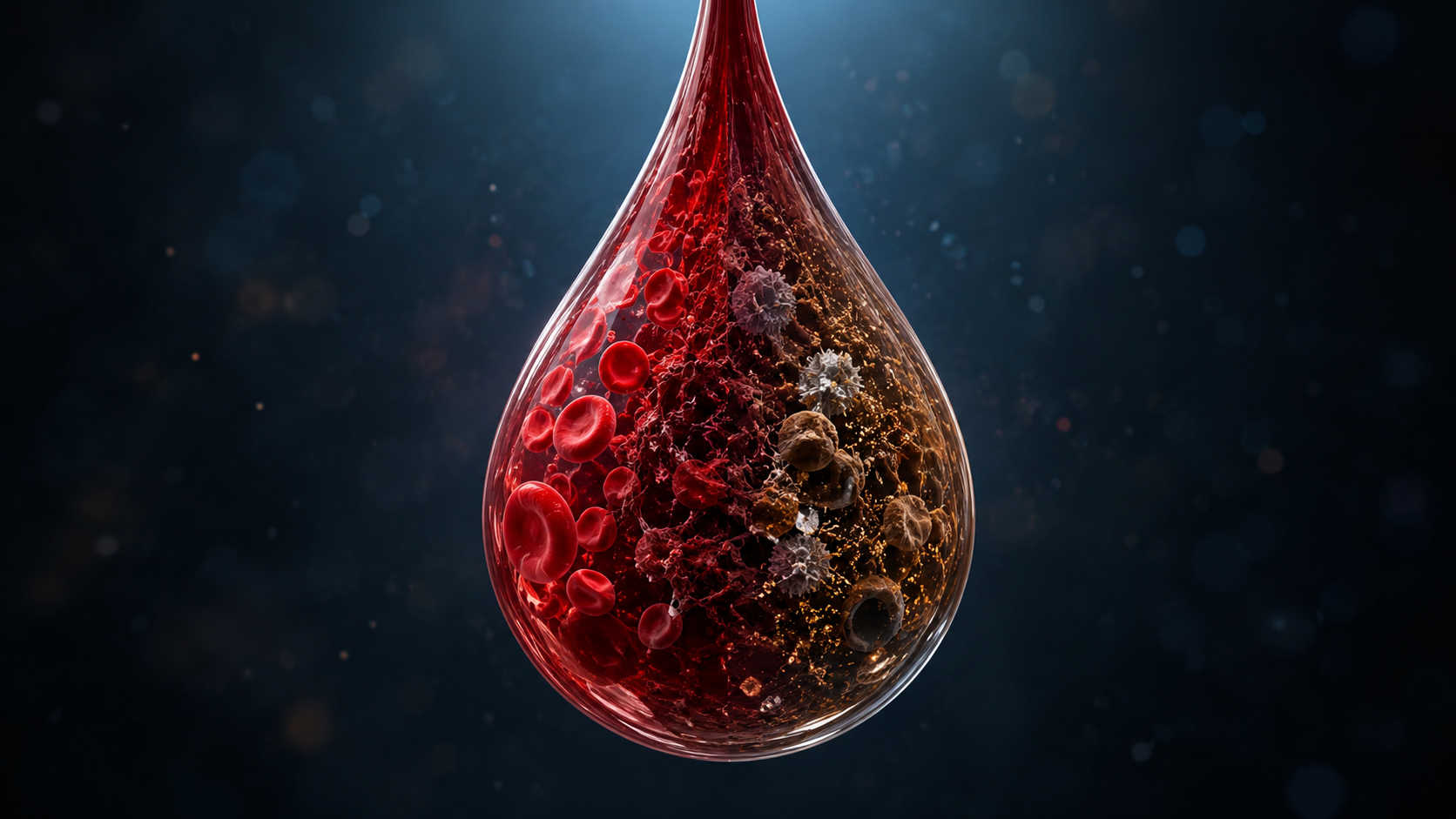
How old is the blood?
A woman presents with vaginal bleeding during pregnancy.
The bleeding may be bright red.
It may be dark red.
It may be brown.
It may be mixed with clots.
It may appear old, thick or fragmented.
One of the most natural questions is:
How old is this blood?
This question sounds simple.
But biologically, it is complex.
Blood does not remain unchanged after it leaves the circulation.
It begins to degrade, oxidize, clot, organize, inflame and interact with surrounding tissues.
A pregnancy bleed may therefore contain a kind of biological timeline.
The challenge is learning how to read it.
This article explores whether we can estimate the age of a pregnancy bleed using forensic principles.
The First Clue: Color
The most obvious sign of blood age is color.
Fresh blood is usually red.
Older blood often becomes darker.
Very old or retained blood may appear brown or almost black.
This happens because hemoglobin changes over time.
Hemoglobin is the oxygen-carrying protein inside red blood cells.
As blood ages, hemoglobin undergoes chemical transformation.
It can oxidize.
It can break down.
Iron can be released.
Pigments can accumulate.
This is why brown pregnancy bleeding is often interpreted as older blood.
But color alone is not enough.
A brown sample may be old.
But how old?
Hours?
Days?
Longer?
To answer that, we need to look deeper.
Blood Is Not a Static Substance
Once blood leaves a vessel, it becomes biologically active.
Several processes begin:
- red blood cells lose integrity
- hemoglobin changes
- clotting begins
- fibrin networks form
- immune cells arrive
- macrophages begin cleanup
- iron is processed
- tissue repair may begin
- RNA and proteins degrade
- bacteria may interact with the sample
- surrounding tissue may contribute additional signals
This means the age of a bleed is not measured by one marker.
It is estimated from a pattern.
The more layers we analyze, the better the reconstruction may become.
Hemoglobin Breakdown
Hemoglobin is one of the most important molecules for estimating blood age.
In fresh blood, most hemoglobin is still inside intact red blood cells.
As blood ages, red blood cells rupture and hemoglobin is released.
This begins a cascade of breakdown.
Possible changes include:
- loss of intact red blood cells
- release of hemoglobin
- formation of oxidized hemoglobin forms
- heme release
- iron release
- pigment formation
In a pregnancy bleeding sample, this may help distinguish:
- very fresh bleeding
- partially degraded bleeding
- old retained bleeding
- hematoma drainage
A fresh red sample may contain many intact erythrocytes.
An older brown sample may contain more degraded cells, free hemoglobin products and pigment.
Oxidation: Why Blood Turns Brown
Oxidation is central to blood aging.
When hemoglobin changes chemically, the color of blood changes.
Bright red blood reflects relatively fresh oxygenated hemoglobin.
Darker blood may reflect deoxygenation and oxidation.
Brown blood often reflects more advanced hemoglobin degradation.
In forensic bloodstain analysis, oxidation and hemoglobin transformation are important tools for estimating time since deposition.
In pregnancy bleeding, the same principle may apply, but the environment is different.
The blood may have been retained inside the uterus, cervix or vagina.
It may have been mixed with mucus, tissue fluid, bacteria or inflammatory cells.
This makes timing more complicated.
But it also makes the sample biologically richer.
Hemosiderin: A Marker of Older Bleeding
One of the clearest signs of older bleeding is hemosiderin.
Hemosiderin is an iron-storage pigment.
It forms when macrophages break down red blood cells and process hemoglobin-derived iron.
This takes time.
Therefore, hemosiderin can suggest that blood has been present long enough for cellular cleanup to begin.
In a bleeding sample, hemosiderin may appear:
- as brown pigment
- inside macrophages
- within tissue fragments
- in areas of old hemorrhage
The presence of hemosiderin does not give an exact timestamp.
But it strongly shifts interpretation away from a purely fresh bleed.
It suggests previous or retained bleeding.
In pregnancy, this may be especially relevant for hematoma-associated bleeding.
Macrophages: The Biological Clock Cells
Macrophages are immune cells that clean up damaged tissue and old blood.
They ingest:
- red blood cell debris
- hemoglobin products
- damaged cells
- tissue fragments
- iron-containing material
Their presence can indicate that the body has already begun responding to the bleeding.
In a very fresh bleed, macrophages may be absent or limited.
In an older bleed, macrophages may be more prominent.
In a resolving hematoma, macrophages may be actively involved in organizing and clearing the old blood.
This makes macrophages a kind of biological clock.
Not a precise clock.
But a sign that time has passed.
Clot Organization
Fresh blood clots differently from old retained blood.
When bleeding occurs, coagulation begins.
Fibrin forms.
Platelets activate.
A clot may develop.
Over time, that clot can change.
It may become:
- more organized
- more compact
- infiltrated by inflammatory cells
- invaded by macrophages
- mixed with tissue fragments
- partially broken down
- attached to surrounding tissue
Microscopy may show fibrin strands or networks.
Older clots may show organization and cellular infiltration.
This is important because pregnancy bleeding may not simply flow out immediately.
It may collect as a hematoma, partially organize, and later drain.
The visible bleeding may therefore be delayed evidence of an earlier event.
RNA Degradation
RNA is more fragile than DNA.
It degrades over time.
This makes RNA potentially useful for estimating sample age, although interpretation is difficult.
In a fresh sample, some RNA signals may still be detectable from intact cells.
In an older sample, RNA may be degraded.
However, some RNA may be protected inside extracellular vesicles.
That means RNA degradation is not simply a clock.
It depends on:
- temperature
- pH
- cell death
- enzymes
- microbial activity
- sample handling
- tissue environment
- extracellular vesicle protection
Still, RNA integrity could become one part of a forensic age-estimation framework.
It may help distinguish recently shed cellular material from older degraded material.
DNA and Protein Degradation
DNA is generally more stable than RNA, but it also changes over time.
Proteins may also degrade, oxidize, fragment or become cross-linked.
Aging blood may show:
- fragmented DNA
- degraded RNA
- oxidized proteins
- altered hemoglobin products
- degraded clot proteins
- modified inflammatory proteins
These molecular degradation patterns could potentially contribute to estimating how long blood has been present.
But again, no single marker is enough.
The best approach is multi-layered.
Fresh Blood, Old Blood and Mixed Blood
A pregnancy bleeding sample may not represent one moment in time.
It may contain layers.
For example:
Fresh bleeding
Possible features:
- bright red color
- many intact red blood cells
- little degradation
- limited macrophage activity
- less hemosiderin
- less clot organization
Old retained bleeding
Possible features:
- brown color
- degraded red blood cells
- hemoglobin breakdown products
- macrophages
- hemosiderin
- fibrin organization
- tissue debris
Mixed bleeding
Possible features:
- brown blood with bright red streaks
- old clots with fresh fluid
- degraded cells plus intact erythrocytes
- signs of both old and new bleeding
Mixed bleeding is especially important.
It may suggest that an older hematoma is draining while new bleeding is also occurring.
This is very different from a single fresh bleeding event.
The Hematoma Timeline
A hematoma is blood that has collected in tissue or between tissue layers.
Once formed, it may evolve.
A possible timeline:
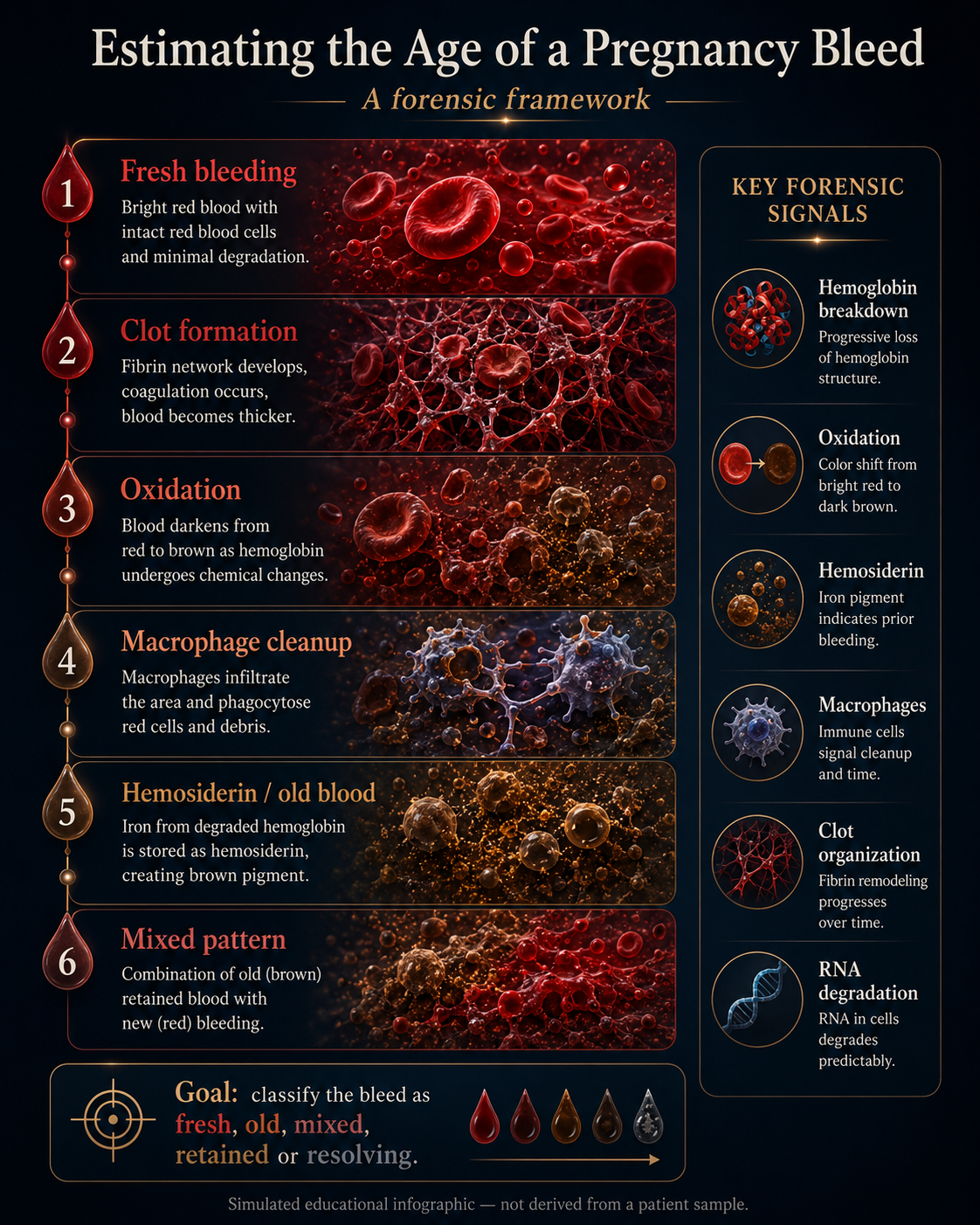
Stage 1: Acute Bleeding
Blood enters a tissue space.
Red blood cells are mostly intact.
Clotting begins.
Stage 2: Early Clot Formation
Fibrin forms.
The blood becomes thicker.
The clot begins to stabilize.
Stage 3: Degradation Begins
Red blood cells rupture.
Hemoglobin is released.
Oxidation begins.
Color darkens.
Stage 4: Inflammatory Cleanup
Macrophages arrive.
Debris is cleared.
Iron is processed.
Hemosiderin may appear.
Stage 5: Organization or Drainage
The hematoma may organize, shrink, persist or drain.
If it drains, the visible bleeding may appear brown, thick or fragmented.
This framework is useful because the bleeding seen outside the body may occur long after the original bleeding event.
Why Exact Timing Is Difficult
It may seem that blood age should be easy to calculate.
But biology is not a controlled laboratory clock.
The rate of blood degradation depends on:
- oxygen exposure
- temperature
- pH
- microbial activity
- tissue contact
- fluid dilution
- clot structure
- immune response
- location of the bleed
- whether blood was retained or immediately expelled
Blood retained in a hematoma may age differently from blood exposed to air.
Blood mixed with vaginal secretions may change differently from blood trapped behind membranes.
Blood in an inflammatory environment may degrade differently from blood in a relatively quiet tissue space.
Therefore, the goal is usually not exact timing.
The goal is classification.
Is the blood likely fresh?
Old?
Repeated?
Mixed?
Retained?
Organizing?
Resolving?
That level of information may already be clinically and scientifically valuable.
The Forensic Age-Estimation Framework
A research framework could combine several layers.
Visual Layer
- bright red
- dark red
- brown
- blackish
- mixed color
- clots
- tissue fragments
Microscopy Layer
- intact red blood cells
- degraded red blood cells
- macrophages
- hemosiderin
- fibrin organization
- tissue debris
Chemistry Layer
- hemoglobin breakdown
- iron release
- oxidation products
- oxidative stress markers
Proteomic Layer
- degraded blood proteins
- clotting proteins
- inflammatory proteins
- tissue repair proteins
RNA Layer
- RNA integrity
- transcript degradation
- microRNA stability
- vesicle-protected RNA
Pattern Layer
- fresh profile
- old profile
- mixed profile
- chronic/recurrent profile
- hematoma drainage profile
This is where AI may eventually become useful.
Not because one marker gives the answer.
But because many weak signals together may form a recognizable pattern.
What Would a Useful Result Look Like?
A future report might not say:
“The blood is exactly 53 hours old.”
Instead, it might say:
Pattern consistent with old retained bleeding, likely days rather than hours, with evidence of macrophage activity, hemoglobin degradation and clot organization. Small fresh component also present.
That kind of interpretation could be much more useful than simply describing the blood as “brown discharge.”
It would convert appearance into biological reconstruction.
Why This Matters
Timing matters because different timelines suggest different biological scenarios.
Fresh active bleeding may suggest one kind of event.
Old brown drainage may suggest another.
Repeated mixed bleeding may suggest a chronic or unstable process.
A resolving hematoma may produce visible bleeding even when the original event occurred earlier.
Without age interpretation, these situations can be collapsed into one category:
“Bleeding.”
But biologically, they may be very different.
The Central Question
The traditional question is:
“Is she bleeding now?”
The forensic timing question is:
“When did this blood first leave the circulation?”
That question changes the entire interpretation.
A woman may present today with bleeding that began biologically days earlier.
The visible event may be late evidence of an earlier tissue event.
That is the core insight.
Conclusion: Reading Time in Blood
Pregnancy bleeding is not only a sign of the present.
It may also carry traces of the past.
Hemoglobin breakdown, oxidation, hemosiderin, macrophages, fibrin organization and RNA degradation may all contribute to estimating the age of a bleed.
No single marker gives perfect timing.
But together, they may reveal whether the bleeding is fresh, old, mixed, repeated or draining from a retained hematoma.
This makes blood-age estimation one of the most fascinating parts of Pregnancy Bleeding Intelligence.
Because the sample may not only tell us where it came from.
It may tell us when the story began.
References and Resource
1. Biphasic Oxidation of Oxy-Hemoglobin in Bloodstains PLoS ONE, July 2011.
A forensic study showing how blood color changes as oxyhemoglobin converts into methemoglobin and hemichrome, explaining why blood can shift from bright red toward dark brown over time.
2. Estimation of the Age of Human Bloodstains Under Simulated Indoor and Outdoor Crime Scene Conditions by ATR-FTIR Spectroscopy Scientific Reports, October 2017.
A study using infrared spectroscopy to estimate bloodstain age under different environmental conditions, highlighting both the potential and the difficulty of forensic blood-age estimation.
3. Short and Long Time Bloodstains Age Determination by Colorimetric Analysis Molecules, October 2021.
A study examining how measurable color changes in bloodstains can be used to estimate time since deposition, based largely on oxidation-related changes.
4. Time Course of Hemosiderin Production by Alveolar Macrophages in a Murine Model Chest, 1999.
An experimental study showing that hemosiderin-loaded macrophages appear after bleeding and persist over time, supporting the idea that hemosiderin can indicate older or previous hemorrhage.
5. Using Total RNA Quality Metrics for Time Since Deposition Estimation of Bloodstains Forensic Science International: Genetics, 2022.
A forensic study exploring RNA degradation as a possible way to estimate the time since a blood sample was deposited.