
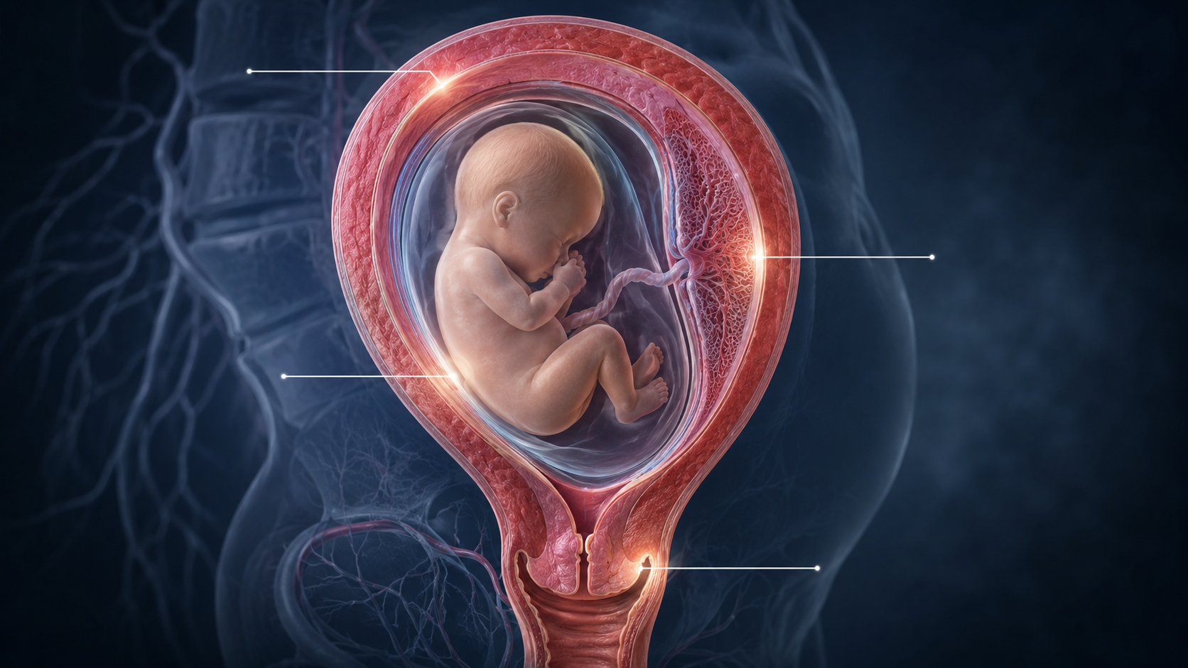
Cervix, decidua, placenta or fetal membranes?
A woman presents with vaginal bleeding during pregnancy.
The first question is often:
How much is she bleeding?
But the deeper forensic question is:
Where did the bleeding come from?
This is not always obvious.
Blood may appear at the vaginal opening, but that does not mean the vagina is the source.
It may have originated from the cervix.
It may have drained from the decidua.
It may have come from the placental edge.
It may have passed through fetal membrane-associated tissue.
It may have started as a hematoma and only become visible later.
In pregnancy bleeding, the final location of the blood is not necessarily the origin of the bleeding.
That is why source reconstruction matters.
This article explores how a forensic framework could help identify whether pregnancy bleeding most likely originated from the cervix, decidua, placenta, fetal membranes or a mixed tissue interface.
The Problem With “Vaginal Bleeding”
The phrase “vaginal bleeding” describes where the blood is seen.
It does not describe where the blood began.
This distinction is important.
A sample may pass through the vagina after originating from:
- cervical tissue
- decidual tissue
- placental-interface tissue
- fetal membranes
- uterine vessels
- a retained hematoma
- a mixed maternal-fetal interface process
Clinically, these may all appear as vaginal bleeding.
Biologically, they may be different events.
A forensic approach therefore asks:
What tissue left evidence in the sample?
Source Attribution
Source attribution means trying to determine the likely tissue of origin.
In pregnancy bleeding, source attribution may rely on multiple evidence layers:
- visual appearance
- timing pattern
- microscopy
- tissue fragments
- microbiome
- chemistry
- proteomics
- DNA/RNA
- methylation
- clinical context
No single layer is enough.
The goal is not to find one perfect marker.
The goal is to combine weak and strong signals into a biological reconstruction.
The Cervix as a Source
The cervix is a common candidate source for bleeding.
During pregnancy, the cervix becomes highly vascular and biologically active.
It may bleed from:
- irritation
- inflammation
- infection
- cervical remodeling
- contact bleeding
- cervical insufficiency
- local tissue fragility
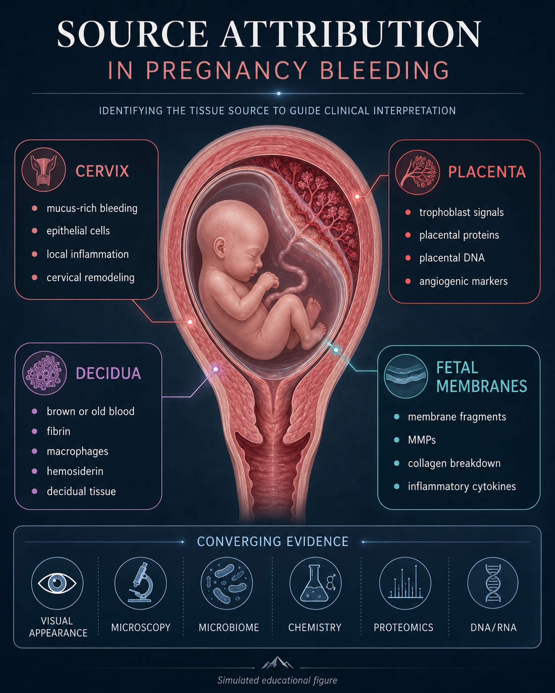
A cervical-source pattern may include:
- mucus-rich bleeding
- blood mixed with cervical secretions
- cervical epithelial cells
- glandular cells
- local inflammatory cells
- cervicovaginal microbiome signatures
- chemokines associated with cervical inflammation
- matrix-remodeling proteins
The cervix is especially interesting because it is not just a passive passageway.
It is a tissue that remodels during pregnancy.
If that remodeling becomes premature, inflammatory or unstable, bleeding may occur as part of a broader biological process.
The Decidua as a Source
The decidua is the pregnancy-modified uterine lining.
It is one of the most important tissues in pregnancy bleeding.
It supports implantation, immune tolerance, vascular adaptation, hemostasis and placental interaction.
A decidual-source pattern may include:
- old or brown blood
- degraded red blood cells
- decidual cells
- decidual tissue fragments
- fibrin
- macrophages
- hemosiderin
- inflammatory proteins
- tissue factor-related signals
- matrix-remodeling markers
- decidual gene-expression or methylation signatures
A decidual bleed may not be a simple surface event.
It may represent disruption at the pregnancy lining.
This is especially relevant for hematoma-like bleeding.
A decidual hematoma may collect blood, age, organize, inflame and later drain.
In that situation, what is seen today may be the delayed result of an earlier decidual event.
The Placenta as a Source
The placenta is another major possible contributor.
Placental involvement may include bleeding near:
- the placental edge
- trophoblast interface
- maternal-fetal boundary
- spiral artery remodeling zones
- placental-decidual attachment area
A placental-source pattern may include:
- trophoblast-derived material
- placental proteins
- placental extracellular vesicles
- placental DNA
- placental methylation patterns
- angiogenic proteins
- vascular stress markers
- placental-interface inflammatory signals
Placental bleeding is not just about blood vessels.
It may reflect abnormal tissue interaction between placenta and decidua.
That makes placental-source reconstruction especially important.
A placental signal in the sample may suggest that the bleeding involved the maternal-fetal interface rather than only the cervix or vagina.
Fetal Membranes as a Source
The fetal membranes are biologically active structures.
They include layers that help contain the amniotic fluid and protect the pregnancy.
Membrane involvement may be relevant when bleeding is associated with:
- inflammation
- infection
- membrane weakening
- tissue remodeling
- preterm labor pathways
- fluid leakage
- ascending infection
A membrane-associated pattern may include:
- membrane-like tissue fragments
- amniotic-associated proteins
- fetal membrane gene-expression patterns
- inflammatory cytokines
- chemokines
- matrix metalloproteinases
- extracellular matrix fragments
- microbiome signatures linked to ascending infection
Membrane-source bleeding may be difficult to identify because the membranes are thin and delicate.
But molecular and protein signals may eventually help reveal membrane stress or involvement.
The Vagina as a Source
The vagina itself can also contribute to bleeding.
A vaginal-source pattern may include:
- squamous epithelial cells
- local mucosal cells
- vaginal microbiome dominance
- local irritation
- trauma-associated bleeding
- inflammatory cells from the vaginal mucosa
However, vaginal-source bleeding must be distinguished from blood that merely passed through the vagina.
This is one reason source reconstruction is difficult.
A sample can collect vaginal cells and microbes during passage even if the original bleeding came from higher up.
That means vaginal signatures may reflect both source and contamination.
The interpretation must be careful.
Mixed Sources
Pregnancy bleeding may not have only one source.
A sample may contain evidence from multiple tissues.
For example:
- old decidual blood draining through the cervix
- placental-interface bleeding mixed with cervical mucus
- hematoma drainage with new fresh bleeding
- membrane inflammation plus decidual bleeding
- cervical bleeding superimposed on old brown hematoma material
This is why source reconstruction should not force a single answer.
A mixed-source model may be more realistic.
The question becomes:
What is the dominant source, and what secondary tissues contributed signals?
Tissue Signatures
Different tissues may leave different signatures.
Cervical Signature
Possible clues:
- mucus
- cervical epithelial cells
- glandular cells
- cervical inflammatory markers
- cervical matrix-remodeling proteins
- cervicovaginal microbiome patterns
Decidual Signature
Possible clues:
- decidual cells
- decidual tissue fragments
- tissue factor
- fibrin
- macrophages
- hemosiderin
- decidual immune proteins
- decidual gene-expression markers
Placental Signature
Possible clues:
- trophoblast material
- placental proteins
- placental DNA
- placental methylation
- angiogenic proteins
- extracellular vesicles
- placental microRNAs
Membrane Signature
Possible clues:
- membrane fragments
- amniotic-associated markers
- MMPs
- collagen fragments
- inflammatory cytokines
- infection-associated signals
- fetal membrane RNA patterns
These signatures are not absolute.
But they provide a framework for thinking.
Biomarker Combinations
The most important principle is combination.
One marker rarely proves the source.
But combinations can become more informative.
Example: Cervical-Dominant Pattern
- mucus-rich bleeding
- cervical epithelial cells
- cervicovaginal microbiome signals
- cervical remodeling proteins
- little placental material
Example: Decidual-Hematoma Pattern
- brown blood
- degraded red blood cells
- macrophages
- hemosiderin
- fibrin
- decidual tissue fragments
- iron-related chemistry
Example: Placental-Interface Pattern
- placental proteins
- trophoblast-derived material
- placental methylation signal
- angiogenic imbalance
- decidual inflammatory markers
Example: Membrane-Stress Pattern
- membrane-associated proteins
- MMPs
- collagen fragments
- inflammatory cytokines
- microbiome signals suggesting ascending inflammation
These are conceptual patterns.
The real power comes from reading them together.
Biological Reconstruction
Biological reconstruction means turning scattered findings into a coherent story.
For example, a report might eventually say:
The bleeding sample contains old degraded blood, macrophage activity, hemosiderin, fibrin organization, decidual tissue signals and minimal fresh blood. Pattern most consistent with drainage from an older decidual hematoma.
Or:
The bleeding sample contains fresh red blood, cervical mucus, cervical epithelial cells, Lactobacillus-dominant microbiome and limited placental markers. Pattern more consistent with cervical-source bleeding.
Or:
The sample contains placental DNA, trophoblast-associated proteins, angiogenic stress markers and decidual inflammation. Pattern suggests placental-decidual interface involvement.
That is the goal of source reconstruction.
Not certainty.
But structured probability.
The Source Reconstruction Matrix
A useful framework could include:
| Evidence layer | What it may contribute |
|---|---|
| Visual appearance | Fresh, old, mixed, clot-rich, mucus-rich |
| Microscopy | Cell types, tissue fragments, inflammation |
| Microbiome | Cervicovaginal ecosystem, infection/dysbiosis |
| Chemistry | Blood age, inflammation, oxidative stress |
| Proteomics | Tissue pathways and protein signatures |
| DNA/RNA | Tissue-of-origin and gene activity |
| Timing pattern | Acute, chronic, recurrent, resolving |
| Clinical context | Gestational age, symptoms, imaging, course |
The source is inferred by convergence.
When multiple independent layers point toward the same tissue, confidence increases.
Why Source Attribution Is Difficult
There are real challenges.
A bleeding sample may be contaminated during passage.
Blood may mix with mucus, vaginal secretions and epithelial cells.
Old and new bleeding may coexist.
Proteins may degrade.
RNA may fragment.
Tissue signatures may overlap.
A single tissue may produce different signals depending on inflammation, gestational age or stress.
Therefore, source reconstruction must be probabilistic.
It should not pretend to offer certainty where none exists.
But uncertainty does not make the approach useless.
It makes structured inference necessary.
Why This Matters
Knowing the source of bleeding could change how we understand risk.
A cervical bleed may mean something different from a decidual hematoma.
A placental-interface bleed may mean something different from vaginal irritation.
A membrane-associated inflammatory bleed may suggest a different pathway than old retained blood draining slowly.
Without source reconstruction, these may all be grouped under the same label:
vaginal bleeding.
But biologically, they may not be the same.
The Central Question
The traditional question is:
“Is there vaginal bleeding?”
The forensic source question is:
“Which tissue system generated the bleeding event?”
That question moves us closer to true Pregnancy Bleeding Intelligence.
Because once we know the likely tissue source, we can begin asking deeper questions:
Why did that tissue bleed?
Was it inflamed?
Was it stressed?
Was it remodeling?
Was it infected?
Was it separating?
Was it healing?
Was the bleeding fresh, old or recurrent?
Source attribution is the doorway to mechanism.
Conclusion: From Blood to Origin
Pregnancy bleeding is often described by appearance and amount.
But a bleeding sample may carry traces of its origin.
The cervix, decidua, placenta and fetal membranes can each leave different biological signatures.
No single finding is enough.
But visual clues, microscopy, microbiology, chemistry, proteomics and DNA/RNA may be combined into a source reconstruction framework.
The aim is not to replace clinical assessment.
The aim is to better read the sample.
Because the blood did not appear from nowhere.
It came from a tissue.
And if we learn to read the traces, pregnancy bleeding may become not just a symptom, but a map back to its biological source.
References and Resource
1. Spatial Transcriptomics of the Fetal Membrane–Decidual Interface Reveals Unique Contributions by Cell Types in Term and Preterm Births PLOS ONE, 19 August 2024.
This study maps the fetal membrane–decidual interface and shows how different cell types contribute to term and preterm birth biology
2. The Maternal-Fetal Interface at Single-Cell Resolution: Uncovering the Complexity of Pregnanc American Journal of Obstetrics & Gynecology, January 2025.
A review of single-cell studies describing the cellular composition and communication networks at the maternal-fetal interface
3. Placental Extracellular Vesicles and Feto-Maternal Communication Frontiers in Immunology, March 2015.
A review explaining how the placenta releases extracellular vesicles carrying proteins, lipids and nucleic acids into maternal circulation.
4. Matrix Metalloproteinase-Induced Cervical Extracellular Matrix Remodelling During Pregnancy and Cervical Cancer Reproduction and Fertility, September 2022.
A review of how MMPs remodel the cervical extracellular matrix during pregnancy, childbirth and pathological tissue change.
5. Matrix Metalloproteinases in the Cervical Mucus Plug in Relation to Gestational Age, Plug Compartment, and Preterm Labor Reproductive Biology and Endocrinology, 24 September 2010.
A study showing that MMPs and TIMPs are present in the cervical mucus plug and may relate to cervical remodeling and preterm labor biology.