
Could Increased Testosterone Drive Endometriosis-Like Inflammation and Miscarriage Risk?
A mechanistic hypothesis linking PCOS, androgen excess, focal tissue activation, progesterone resistance and pregnancy-interface instability
Article type
Hypothesis review / applied research model
Introduction
Endometriosis, PCOS, infertility, implantation failure and miscarriage are usually discussed as separate clinical problems.
PCOS is often framed as an ovarian and metabolic condition.
Endometriosis is often framed as an estrogen-dependent inflammatory disease.
Miscarriage is often framed through embryo genetics, uterine anatomy, thrombophilia, immune factors, endocrine support or chance.
Progesterone treatment is often framed as either sufficient or unnecessary after placental takeover.
But some patients do not fit neatly into these categories.
A woman may have PCOS, androgenic symptoms, low fertility, suspected or confirmed endometriosis, strong cyclic symptoms, mood and behavioral androgen sensitivity, IVF history, pregnancy loss or pregnancy instability after progesterone withdrawal. She may not have severe virilization. She may not have a deep voice, permanent clitoral enlargement or male-pattern scalp hair loss. Yet she may still have clinically relevant hyperandrogenism.
The question becomes:
Could increased testosterone or androgen excess act as an upstream driver of endometriosis-like focal inflammation, progesterone resistance and miscarriage risk in a subgroup of patients?
This article explores that hypothesis.
It is not a claim that testosterone universally causes endometriosis. It is not a claim that every miscarriage in PCOS is androgen-driven. It is not a claim that all women with high testosterone require suppression during pregnancy.
It is a mechanistic framework.
The core idea is this:
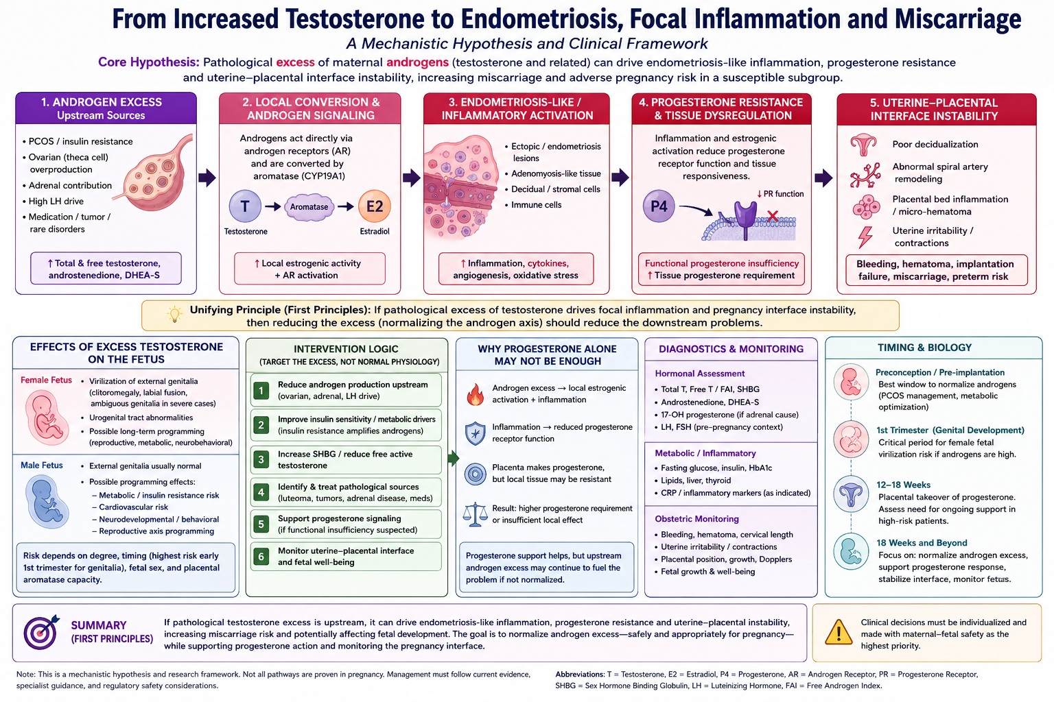
In susceptible patients, pathological androgen excess may contribute to focal uterine, decidual, placental-bed or endometriosis-like inflammatory activation. This may occur through local aromatization to estradiol, androgen receptor signaling, insulin-driven ovarian androgen production, altered progesterone responsiveness, immune activation, vascular instability and tissue-specific inflammatory hotspots. In pregnancy, this may create a state where placental progesterone production is normal, but local tissue response remains insufficient.
The endpoint could be implantation failure, bleeding, hematoma, uterine-interface instability, recurrent pregnancy loss or miscarriage in a subgroup.
This is speculative. But it is biologically plausible enough to discuss seriously.
The first-principles argument
The starting point is simple.
If an abnormal excess of testosterone is causally driving focal inflammation, tissue instability or pregnancy-interface dysfunction, then the rational biological objective is to remove or normalize the excess.
Not to block normal fetal androgen physiology.
Not to suppress all androgen signaling.
Not to treat testosterone as inherently bad.
The objective would be:
Normalize pathological androgen excess while preserving normal maternal, placental and fetal physiology.
This distinction matters.
Normal testosterone has biological roles.
Pathological excess may create abnormal signaling.
Pregnancy requires hormonal balance, not hormonal excess.
If testosterone is upstream of the tissue problem, then progesterone alone may be a downstream stabilizer rather than a root-cause treatment. Progesterone support may help calm inflammatory activation, stabilize the uterine interface or compensate for functional progesterone insufficiency. But if androgen excess continues to feed local estrogenic or inflammatory activation, progesterone may not fully solve the problem.
In that model, the treatment question becomes:
Can we identify, measure and safely normalize abnormal androgen signaling before or during pregnancy in the subgroup where it matters?
The proposed chain
The hypothesis can be expressed as a pathway:
PCOS or hyperandrogenic biology
→ increased total or free testosterone, androstenedione, DHEA-S or related androgens
→ insulin resistance, low SHBG and amplified ovarian/adrenal androgen production
→ local androgen receptor effects and/or aromatization to estradiol
→ focal estrogenic and inflammatory activation in susceptible tissue
→ endometriosis-like lesion activity, adenomyosis-like uterine tissue response or decidual/placental-bed inflammation
→ progesterone resistance or increased tissue progesterone requirement
→ impaired decidualization, vascular instability, bleeding, hematoma, uterine irritability or placental-interface dysfunction
→ implantation failure, pregnancy loss, miscarriage or later pregnancy complications in a subgroup
This does not need to happen uniformly across the uterus.
The disease signal may be spatial.
One region may be stable.
Another region may have inflammatory activation.
A third region may have higher aromatase activity.
A fourth region may be more progesterone-resistant.
A small hotspot may contribute more to risk than a broad low-grade background signal.
This is why a patient may have normal-looking systemic blood results but still have a localized tissue problem.
Why PCOS matters in this hypothesis
PCOS is not only an ovulation disorder.
It is commonly associated with androgen excess, insulin resistance, metabolic dysfunction, irregular ovulation, altered endometrial receptivity, low fertility and increased pregnancy risks in some populations.
In PCOS, androgen excess can come from several interacting systems:
-
ovarian theca-cell androgen production
-
increased LH drive
-
insulin amplification of androgen production
-
reduced SHBG leading to higher free androgen exposure
-
adrenal androgen contribution
-
obesity or metabolic inflammation in some patients
-
genetic and epigenetic susceptibility
This means that a woman with PCOS may have more than high serum testosterone. She may have a whole endocrine-metabolic tissue environment that changes how the endometrium, decidua, immune system and placenta behave.
The question is not only:
Is testosterone high?
The better question is:
Is androgen signaling biologically excessive in the relevant tissue environment?
That may depend on free testosterone, SHBG, insulin, aromatase activity, androgen receptors, estrogen receptors, progesterone receptors, inflammatory cytokines and local tissue history.
Testosterone is not only testosterone: local conversion matters
A major mechanism in this hypothesis is aromatization.
Testosterone can be converted into estradiol by aromatase. Estradiol is a key driver of estrogen-dependent gynecological tissue activity. Endometriosis is often described as estrogen-dependent, inflammatory and progesterone-resistant.
So a high androgen substrate state could theoretically feed local estrogenic activation if the tissue expresses aromatase.
The possible pathway is:
high testosterone or androstenedione
→ local aromatase activity
→ increased local estradiol
→ estrogen receptor activation
→ inflammatory cytokines, angiogenesis and tissue remodeling
→ endometriosis-like lesion persistence or focal decidual instability
This is important because serum estrogen measurements may not reflect local estrogen production.
A woman may not have globally excessive estrogen, but a focal tissue region could still experience abnormal estrogenic activation.
That is the “geographical hotspot” concept.
Androgen receptors may also matter directly
The hypothesis should not rely only on aromatization.
Androgens can also signal through androgen receptors in reproductive tissues. Androgen receptor expression exists in endometrial tissue and may influence stromal and epithelial behavior.
This creates several possibilities:
-
Androgens may support normal endometrial function in some contexts.
-
Androgens may increase progesterone receptor expression in some experimental settings.
-
Androgen excess may disrupt receptivity or contribute to abnormal tissue behavior in other contexts.
-
The effect may depend on dose, timing, receptor distribution and inflammatory state.
This is why the hypothesis must remain nuanced.
Androgens are not always harmful.
Low androgen signaling may also be relevant in some endometriosis pain models.
But pathological androgen excess in a PCOS-like pregnancy or fertility context may still be abnormal.
The key is not testosterone alone.
The key is androgen signaling in a susceptible tissue system.
Endometriosis as focal tissue disease
Endometriosis is not simply “uterine lining outside the uterus.”
It is a complex inflammatory and endocrine tissue disease. It involves ectopic endometrial-like tissue, immune dysfunction, angiogenesis, fibrosis, pain signaling, altered steroid metabolism, local estrogen production and progesterone resistance.
This is highly compatible with a focal activation model.
Endometriosis lesions are not evenly distributed. They may differ in activity, depth, vascularity, innervation, inflammatory signaling, steroid receptor expression and response to treatment.
Some lesions may be relatively inactive.
Some may be inflammatory.
Some may be estrogen-sensitive.
Some may be progesterone-resistant.
Some may interact with adjacent tissues, nerves, vessels or immune cells.
If testosterone excess contributes to local estrogenic or androgenic activation, the effect may not be visible as a uniform systemic signal. It may show up as focal pain, bleeding, implantation dysfunction, placental-bed instability or miscarriage risk.
Progesterone resistance: the critical bridge
Progesterone is central to implantation, decidualization, immune tolerance, uterine quiescence and pregnancy maintenance.
But endometriosis is associated with progesterone resistance.
Progesterone resistance means that the tissue does not respond normally to progesterone. This can involve altered progesterone receptor expression, receptor isoform imbalance, inflammatory signaling, epigenetic changes, estrogen dominance and local immune activation.
This is one of the most important bridges between endometriosis and miscarriage risk.
If a tissue region is progesterone-resistant, then normal progesterone levels may not produce normal progesterone effects.
The patient may not have an absolute progesterone deficiency.
She may have a functional progesterone insufficiency.
That means:
Progesterone is present, but tissue response is inadequate.
This can explain why some patients deteriorate when progesterone is stopped after 12 weeks, even though the placenta is producing progesterone.
The placenta may be doing its job in the average sense.
But the local tissue may require more progesterone signal or may not respond properly to normal levels.
Why stopping progesterone at 12 weeks may not work for all patients
In standard IVF and early pregnancy care, progesterone support is often stopped around the time placental progesterone production becomes established. Biologically, this makes sense for many patients.
But not all patients are average.
Some patients may have:
-
endometriosis
-
adenomyosis
-
PCOS
-
hyperandrogenism
-
previous miscarriage
-
bleeding episodes
-
hematoma
-
inflammatory pelvic disease
-
recurrent implantation failure
-
progesterone resistance
-
abnormal uterine tissue response
-
placental-bed vulnerability
In these patients, the question is not only:
Can the placenta produce progesterone?
It is:
Is placental progesterone sufficient for this patient’s tissue state?
If the problem is focal inflammation, androgen-driven local estrogenic activation or progesterone resistance, stopping progesterone at 12 weeks may remove a stabilizing force before the uterine-interface problem is fully controlled.
This does not prove that all such patients need progesterone throughout pregnancy. But it supports the idea that the standard stop point may not fit every subgroup.
The pregnancy phase after 18 weeks
From around mid-pregnancy onward, the endocrine system is dominated by the placenta.
Progesterone is high. Estrogen is rising. The fetus, placenta and maternal endocrine systems are tightly integrated.
If a patient has excessive maternal androgen exposure at this stage, several questions matter:
-
Is total testosterone high?
-
Is free testosterone high?
-
Is SHBG low?
-
Is androstenedione elevated?
-
Is DHEA-S elevated?
-
Is the source ovarian, adrenal, placental, metabolic or medication-related?
-
Is the placenta converting androgens normally?
-
Is the fetus exposed?
-
Is there bleeding, hematoma, placental-bed dysfunction or fetal growth issue?
-
Is there evidence of gestational diabetes, preeclampsia risk or inflammation?
At this stage, the concern is not only miscarriage. It may include placental function, fetal growth, maternal metabolic complications and developmental programming.
The hypothesis after 18 weeks becomes:
Pathological maternal androgen excess may sustain focal uterine-interface inflammation and functional progesterone insufficiency, even in the presence of high placental progesterone.
Could excess testosterone affect the fetus?
Yes, if exposure is high enough, timing is critical and placental buffering is insufficient.
For a female fetus, the clearest established risk of significant androgen excess is virilization, especially if exposure occurs during early genital development. This can include clitoral enlargement, labial fusion or ambiguous genitalia in severe cases.
For a male fetus, the genital effects may be less obvious because male genital development normally depends on androgen signaling. The concern is more about developmental programming: metabolic, cardiovascular, reproductive-axis or neurobehavioral effects.
This distinction matters.
The argument for normalizing excess testosterone is not only about maternal inflammation or miscarriage risk. It may also be about preventing abnormal fetal androgen exposure.
However, the goal must be precise.
The goal is not to suppress normal fetal androgen physiology.
The goal is to reduce pathological maternal androgen excess toward physiological range.
That is a very different concept from crude anti-androgen blockade.
Why “blocking testosterone” is not the correct wording
A key conceptual correction is needed.
The intervention target should not be described as “blocking testosterone” in a broad sense.
The better wording is:
normalize pathological androgen excess
or
reduce abnormal free androgen exposure
or
remove the pathological upstream androgen driver
This matters because normal androgen signaling is part of fetal and maternal physiology. But excess androgen exposure may be harmful.
So the therapeutic concept should be:
target the abnormal differential increase, not baseline physiology.
That may include reducing insulin-driven ovarian androgen production, increasing SHBG, identifying an ovarian/adrenal source, treating a pathological luteoma or tumor if present, stabilizing metabolic drivers, or using pregnancy-compatible strategies that reduce androgen excess without harming fetal development.
Possible sources of androgen excess
A serious model must distinguish causes.
PCOS-related androgen excess
This is the most common context. It may involve ovarian androgen overproduction, insulin resistance, low SHBG and metabolic inflammation.
Adrenal contribution
DHEA-S elevation may suggest adrenal involvement. Non-classic congenital adrenal hyperplasia may be relevant in selected cases.
Ovarian hyperreactivity
Some ovarian states may produce high androgen levels. In pregnancy, rare conditions such as luteoma of pregnancy or hyperreactio luteinalis can produce significant androgen excess.
Medication or supplement exposure
Testosterone therapy, DHEA, anabolic agents or “hormone optimization” products can contribute.
Assay artifact
Pregnancy changes binding proteins and hormone assays can be misleading. Total testosterone alone may not be enough.
Placental handling
Placental aromatase normally converts androgens into estrogens. If placental capacity is overwhelmed or abnormal, fetal and maternal effects may differ.
What should be measured?
If this hypothesis is to become useful, it must become measurable.
A possible diagnostic framework includes:
Androgen profile
-
total testosterone
-
free testosterone
-
calculated free androgen index
-
SHBG
-
androstenedione
-
DHEA-S
-
17-OH progesterone when adrenal contribution is suspected
-
possibly 11-oxygenated androgens in research settings
Metabolic profile
-
fasting glucose
-
fasting insulin
-
HbA1c
-
lipid profile
-
liver markers
-
BMI and body composition where relevant
-
gestational diabetes screening
Inflammation and tissue-state context
-
CRP where relevant
-
white blood cell context
-
infection evaluation if symptoms suggest it
-
endometriosis history
-
adenomyosis history
-
pelvic pain pattern
-
bleeding episodes
-
hematoma history
Pregnancy interface monitoring
-
ultrasound
-
hematoma presence or resolution
-
cervical length
-
placental position and morphology
-
fetal growth
-
uterine artery Doppler where indicated
-
blood pressure
-
symptoms of uterine irritability or contractions
Progesterone response context
-
progesterone treatment history
-
symptoms after progesterone withdrawal
-
bleeding after withdrawal
-
response to reintroduction
-
prior pregnancy outcomes with and without extended progesterone
-
evidence of progesterone resistance or endometriosis
The goal is not to test everything randomly.
The goal is to determine whether a patient belongs to a biologically coherent subgroup.
What would support the hypothesis?
The hypothesis would become more convincing if research found patterns such as:
-
PCOS or hyperandrogenic patients with endometriosis-like symptoms have higher miscarriage risk than non-hyperandrogenic endometriosis patients.
-
Free testosterone or low SHBG predicts bleeding, hematoma, implantation failure or pregnancy loss.
-
Androgen excess correlates with inflammatory markers or tissue-state markers.
-
Endometriosis lesions in affected patients show aromatase activity and androgen receptor activation.
-
Patients who worsen after progesterone withdrawal have higher androgen activity or lower progesterone response markers.
-
Normalizing androgen excess before embryo transfer improves implantation or reduces loss.
-
Continuing progesterone helps only partially unless androgen-metabolic drivers are corrected.
-
Placental markers differ in PCOS-hyperandrogenic pregnancies with complications.
-
Fetal outcomes differ by maternal free androgen exposure.
These are testable questions.
What would argue against the hypothesis?
The model must include counterarguments.
Testosterone may suppress cycling
In some people, high androgen exposure may suppress menstruation and ovarian cycling. This could reduce endometriosis symptoms in certain contexts.
Low androgen models exist
Some literature suggests low androgen tone may contribute to pain sensitivity or endometriosis-related symptoms. This argues against a simple “high testosterone causes endometriosis” model.
PCOS and endometriosis are often different phenotypes
PCOS and endometriosis are traditionally viewed as distinct reproductive disorders. Their coexistence is possible, but the mechanisms may not be shared in every patient.
Pregnancy often suppresses classic endometriosis
Pregnancy often reduces cycling and menstruation, which may reduce classic endometriosis activity. But this does not necessarily eliminate focal inflammatory or decidualized lesion behavior.
Hyperandrogenism does not always predict miscarriage
Some studies show associations between PCOS, hyperandrogenism and pregnancy complications, but miscarriage-specific links are inconsistent.
Progesterone resistance may be independent
Progesterone resistance in endometriosis may occur independently of testosterone excess. Androgen excess may be a contributor in some patients, not the universal cause.
These counterarguments do not destroy the hypothesis. They define its boundaries.
The correct model is subgroup-specific.
A more precise subgroup hypothesis
The broad claim “testosterone causes endometriosis and miscarriage” is too crude.
A better hypothesis is:
In a subgroup of patients with PCOS or hyperandrogenic biology, pathological free androgen excess may act as an upstream amplifier of endometriosis-like focal inflammation, local estrogenic activity and progesterone resistance. This may create uterine-interface instability that contributes to implantation failure, bleeding, hematoma, pregnancy loss or miscarriage, especially when progesterone support is withdrawn after placental transition.
That is much more defensible.
What could be done before conception or embryo transfer?
The best intervention window is likely before pregnancy or before embryo transfer.
Possible strategies to investigate:
1. Phenotype the patient
Identify PCOS, hyperandrogenism, suspected endometriosis, adenomyosis, previous miscarriage, IVF failure, inflammatory symptoms and metabolic risk.
2. Normalize metabolic drivers
If insulin resistance is driving ovarian androgen production, metabolic treatment may reduce androgen excess more safely than direct anti-androgen drugs.
This may include:
-
nutrition strategy
-
exercise where appropriate
-
weight optimization when relevant
-
metformin in selected PCOS patients
-
inositol in selected contexts
-
glucose and insulin monitoring
3. Reduce free androgen exposure
Because free testosterone may matter more than total testosterone, strategies that raise SHBG or reduce insulin resistance may lower active androgen exposure.
4. Avoid transferring into an activated tissue state
In IVF, freeze-all strategies or delayed transfer may allow time to optimize the endocrine-metabolic-inflammatory environment.
5. Suppress endometriosis or adenomyosis before transfer
Some patients may benefit from GnRH agonist or antagonist downregulation before embryo transfer, especially in endometriosis or adenomyosis contexts. This does not specifically target testosterone but may reduce broad ovarian steroid activity and tissue activation.
6. Treat visible pathology
Endometrioma, adenomyosis, fibroids, polyps or severe lesions may require individualized specialist management.
7. Define progesterone strategy before transfer
If a patient has suspected progesterone resistance or prior loss after progesterone withdrawal, the progesterone plan should be deliberate rather than automatic.
What could be done during pregnancy?
During pregnancy, the intervention logic changes.
The goal remains normalization of pathological androgen excess, but treatment options are more constrained because fetal development must be protected.
Possible approaches to investigate include:
1. Confirm that androgen excess is real
Pregnancy hormone measurement is complex. Total testosterone alone can mislead. Free androgen index, SHBG and androgen source matter.
2. Identify dangerous causes
Very high testosterone, rapid virilization or severe symptoms should prompt evaluation for ovarian or adrenal sources, luteoma, hyperreactio luteinalis, tumor or medication exposure.
3. Treat metabolic drivers
If androgen excess is PCOS-metabolic driven, glucose control, insulin resistance management and gestational diabetes prevention become upstream androgen strategies.
4. Continue progesterone if there is a clinical pattern
If progesterone withdrawal triggers bleeding, uterine symptoms or instability, extended progesterone support may be rational in selected patients.
This should be framed as support for functional progesterone insufficiency or inflammatory uterine-interface instability, not as proof of absolute progesterone deficiency.
5. Monitor placenta and uterine interface
Pregnancy surveillance should focus on:
-
bleeding
-
hematoma
-
cervical length
-
placental findings
-
fetal growth
-
Doppler patterns where relevant
-
blood pressure
-
glucose regulation
-
symptoms of uterine irritability
6. Avoid crude anti-androgen therapy
Broad androgen receptor blockade or drugs known to interfere with fetal androgen physiology are generally problematic in pregnancy. The goal is not to block normal androgen signaling, but to identify safe ways to normalize pathological excess.
7. Consider research-level biomarkers
Future research may identify tissue-state markers, inflammatory signatures or placental markers that better identify the subgroup.
Why progesterone alone may not be enough
Progesterone may help, but it may not fully solve the upstream problem.
If the pathway is:
androgen excess → local estrogenic/inflammatory activation → progesterone resistance
then progesterone is treating the downstream insufficiency.
It may be necessary.
It may reduce risk.
It may stabilize the pregnancy interface.
But it may not remove the upstream fuel.
This could explain why some women need progesterone longer than expected, or why progesterone helps but does not fully prevent complications.
A more complete model would combine:
-
androgen normalization where safe
-
metabolic stabilization
-
inflammation assessment
-
progesterone support where indicated
-
placental and uterine-interface monitoring
Could the placenta simply convert testosterone to estrogen anyway?
The placenta normally aromatizes androgen precursors into estrogens.
This is usually protective because it helps prevent excessive androgen exposure to the fetus. But in this model, the same pathway can create another problem:
high androgen substrate → increased estrogenic output or local estrogenic activation
The issue is not necessarily systemic estrogen levels.
The issue is local estrogenic signaling in a susceptible inflammatory tissue environment.
This is why the model is not:
testosterone is bad
It is:
excess androgen substrate may feed abnormal local steroid signaling in susceptible tissue.
Spatial model: hotspots rather than global inflammation
A useful way to think about this is spatially.
Imagine the reproductive tissue system as a map.
Most regions may be stable.
Some regions may have mild low-grade inflammation.
A few regions may be high-activation hotspots.
A hotspot could have:
-
high aromatase
-
high inflammatory cytokines
-
altered progesterone receptors
-
vascular instability
-
immune activation
-
lesion persistence
-
local estrogen sensitivity
-
tissue remodeling
The total pregnancy risk may depend less on the average inflammatory level and more on the weighted effect of hotspots.
A small but intense lesion near a vulnerable uterine or placental interface may matter more than broad mild inflammation elsewhere.
This is the logic behind the focal inflammation model.
How this connects to miscarriage
Miscarriage has many causes. Embryo chromosomal abnormalities are common, especially in early miscarriage. Uterine anatomy, thrombophilia, infection, endocrine disease, autoimmune disease, placental factors and chance may all matter.
This model does not replace those explanations.
It adds a subgroup pathway:
androgen excess and PCOS-like metabolic biology may contribute to a tissue environment that is less tolerant of pregnancy, less responsive to progesterone and more prone to focal inflammatory instability.
Possible miscarriage-related mechanisms include:
-
impaired implantation
-
abnormal decidualization
-
impaired spiral artery remodeling
-
placental-bed inflammation
-
hematoma formation
-
uterine irritability
-
cervical or myometrial inflammatory signaling
-
progesterone resistance
-
local estrogen excess
-
immune imbalance
-
oxidative stress
This is especially relevant for patients with overlapping PCOS, endometriosis, recurrent loss, IVF history, bleeding episodes or progesterone-withdrawal sensitivity.
What kind of study could test this?
A serious research program could include:
Cohort 1: PCOS IVF patients
Compare hyperandrogenic and non-hyperandrogenic PCOS patients undergoing frozen embryo transfer.
Measure androgen profile before transfer and during pregnancy.
Track implantation, bleeding, hematoma, miscarriage, progesterone use and live birth.
Cohort 2: Endometriosis IVF patients
Stratify by androgen profile, SHBG, insulin resistance and inflammatory markers.
Assess whether hyperandrogenic endometriosis patients have different outcomes.
Cohort 3: Patients who worsen after progesterone withdrawal
Identify patients who develop bleeding, uterine symptoms or pregnancy instability after stopping progesterone around 10–12 weeks.
Compare androgen markers, PCOS features, endometriosis symptoms and outcomes.
Tissue studies
In endometrial biopsy, decidual tissue or surgical endometriosis samples, measure:
-
aromatase
-
androgen receptor
-
estrogen receptors
-
progesterone receptor isoforms
-
inflammatory cytokines
-
immune cell markers
-
angiogenesis markers
-
fibrosis/remodeling markers
Spatial modeling
Map lesion activity and inflammation as weighted tissue signals rather than one global diagnosis.
This could connect imaging, symptoms, biomarkers and outcomes.
Clinical caution
This hypothesis must not be translated into unsupervised treatment.
No pregnant patient should attempt to lower testosterone with unapproved medication, supplements or anti-androgenic drugs without specialist care.
This is especially important because:
-
fetal androgen physiology matters
-
pregnancy hormone interpretation is complex
-
anti-androgen drugs may be unsafe
-
miscarriage has many causes
-
endometriosis treatment in pregnancy is limited
-
PCOS pregnancies need individualized metabolic care
-
high testosterone can sometimes indicate rare serious conditions
The safe clinical message is:
If androgen excess is suspected in pregnancy, measure it properly, identify the source, assess maternal-fetal risk, and manage through a specialist team.
What this hypothesis does well
It connects several observations that are often separated:
-
PCOS can involve androgen excess.
-
PCOS can involve low fertility and pregnancy complications.
-
Endometriosis is inflammatory, estrogen-responsive and progesterone-resistant.
-
Testosterone can be converted to estradiol.
-
Pregnancy progesterone production may not guarantee adequate local progesterone response.
-
Some patients worsen after progesterone withdrawal.
-
Focal tissue activity may matter more than global hormone levels.
-
Excess androgens may affect fetal development or programming.
-
Normalizing pathological excess is a rational first-principles objective.
This makes the model worth exploring.
What this hypothesis does not prove
It does not prove that testosterone causes endometriosis.
It does not prove that testosterone causes miscarriage.
It does not prove that all PCOS patients need androgen-lowering treatment.
It does not prove that progesterone should be continued throughout pregnancy for all high-risk patients.
It does not prove that anti-androgen drugs should be used during pregnancy.
It does not prove that every focal inflammation pattern is androgen-driven.
It is a research framework.
Balanced conclusion
The hypothesis can be stated clearly:
In a susceptible subgroup, pathological androgen excess may contribute to endometriosis-like focal inflammation, local estrogenic activation, progesterone resistance and uterine-interface instability. This may increase the risk of implantation failure, bleeding, hematoma, miscarriage or pregnancy complications, especially in women with PCOS, hyperandrogenism, endometriosis symptoms, IVF history or progesterone-withdrawal sensitivity.
The first-principles logic is strong:
If pathological testosterone excess is upstream, the rational objective is to normalize the excess.
But the clinical implementation must be careful:
Normalize abnormal androgen signaling without disrupting normal fetal and placental physiology.
The most promising intervention window is likely before conception or before embryo transfer, when PCOS, insulin resistance, free androgen exposure, endometriosis activity and uterine tissue state can be optimized more safely.
During pregnancy, the focus should shift to confirming androgen excess, identifying its source, managing metabolic drivers, supporting progesterone response where clinically justified, and monitoring the placenta–decidua–uterine interface.
This is not standard doctrine yet.
But it is a coherent hypothesis.
It deserves structured research because it may explain a subgroup of patients who are currently told that “the placenta should be doing the job” when their tissue biology may be telling a more complicated story.
Referenses
Pathogenesis-Based Diagnosis and Treatment of Endometriosis
NIH PMC
Review discussing endometriosis as a complex disease involving lesion variability, aromatase activity, progesterone resistance, inflammation, angiogenesis, immunologic changes and fibrosis. This supports the idea that endometriosis is not a uniform systemic condition but a tissue-level disease with variable lesion biology.
Inflammatory Status Influences Aromatase and Steroid Receptor Expression in Endometriosis
NIH PMC
Study showing increased aromatase expression in eutopic endometrium from endometriosis patients compared with controls, supporting the biological plausibility of local steroid conversion and estrogenic activation in endometriosis tissue environments.
Progesterone Resistance in Endometriosis: Current Evidence and Putative Mechanisms
International Journal of Molecular Sciences / MDPI
Review describing progesterone resistance as impaired endometrial response to progesterone and summarizing evidence that loss of progesterone signaling contributes to endometriosis persistence and treatment failure.
Progesterone Resistance, Aromatase, and Inflammation: The Important Triad in Endometriosis
Springer
Review connecting reduced progesterone responsiveness with inflammation and aromatase biology in endometriosis. Useful for supporting the triad of local estrogenic signaling, inflammation and impaired progesterone response.
Androgen Excess: A Hallmark of Polycystic Ovary Syndrome
Frontiers in Endocrinology
Review of androgen excess in PCOS, including androgen synthesis, androgen receptor signaling, 11-oxygenated androgens and therapeutic approaches targeting hyperandrogenism. Supports the PCOS/hyperandrogenic starting point of the hypothesis.
Sustained Maternal Hyperandrogenism During PCOS Pregnancy Reduced by Metformin in Non-Obese Women Carrying a Male Fetus
Journal of Clinical Endocrinology & Metabolism / NIH PMC
Study describing androgen patterns during pregnancy in women with PCOS and controls, and evaluating metformin effects. Relevant to whether androgen excess can persist during pregnancy.
The Impact of Hyperandrogenemia on Pregnancy Complications and Outcomes in Patients with PCOS
Journal of Maternal-Fetal & Neonatal Medicine / Taylor & Francis
Meta-analysis reporting that hyperandrogenism in PCOS is associated with higher risks of gestational diabetes and preeclampsia, while not showing significant differences for several other outcomes including miscarriage in that analysis. Useful both as support and limitation.
Pre-Conception Androgen Levels and Obstetric Outcomes in Polycystic Ovary Syndrome
NIH PMC
Study addressing the relationship between pre-pregnancy androgen levels and obstetric/neonatal outcomes in PCOS, emphasizing that the link between hyperandrogenism and adverse outcomes is not fully understood.
Hyperandrogenism and Its Possible Effects on Endometrial Receptivity: A Review
International Journal of Molecular Sciences / MDPI
Review focusing on androgen excess, endometrial receptivity, infertility and pregnancy loss mechanisms. Particularly relevant to the bridge between androgen excess and impaired implantation or reproductive outcomes.
Androgens, Endometriosis and Pain
Frontiers in Reproductive Health
Important counterbalancing review. Discusses evidence that low androgen tone may contribute to endometriosis-related pain and that androgens may sometimes reduce inflammation. This should be included to show that the testosterone–endometriosis relationship is not one-directional or settled.
Endocrine Aspects of Endometriosis
European Journal of Endocrinology
Recent endocrine review noting emerging evidence that lower systemic bioavailable testosterone may play a role in endometriosis development or symptom severity, while also emphasizing estrogen-driven lesion survival and local steroidogenesis. Important counterargument to a simplistic high-testosterone model.
Gestational Hyperandrogenism in Developmental Programming
Endocrinology / Oxford Academic
Review of causes of gestational hyperandrogenism and its possible effects on fetal developmental trajectory and offspring outcomes. Supports discussion of fetal exposure and developmental programming.
Maternal Hyperandrogenemia and the Long-Term Neuropsychological, Sex-Specific and Metabolic Development of Children
International Journal of Molecular Sciences / MDPI
Review discussing possible long-term offspring effects of elevated maternal androgen levels, including neurodevelopmental, metabolic and behavioral outcomes. Relevant to fetal and child outcome considerations.