
Can proteins tell us which tissue is involved?
Pregnancy bleeding can be seen with the eye.
It can be examined under a microscope.
It can be studied for microbes.
It can be analyzed chemically.
But proteins add another layer.
Proteins are the working molecules of biology.
They build tissue.
They signal inflammation.
They regulate blood vessels.
They remodel membranes.
They control clotting.
They reflect placental activity.
They respond to injury.
If DNA is the blueprint, proteins are much closer to the actual biological event.
This makes proteomics — the large-scale study of proteins — especially interesting for pregnancy bleeding.
The question becomes:
Can proteins in a bleeding sample help reveal which tissues were involved and what biological process was active?
From Blood to Tissue Interface
A pregnancy bleeding sample may not come from only one source.
It may contain signals from:
- maternal blood
- cervix
- vagina
- decidua
- placenta
- fetal membranes
- immune cells
- blood vessels
- clotting systems
- damaged tissue
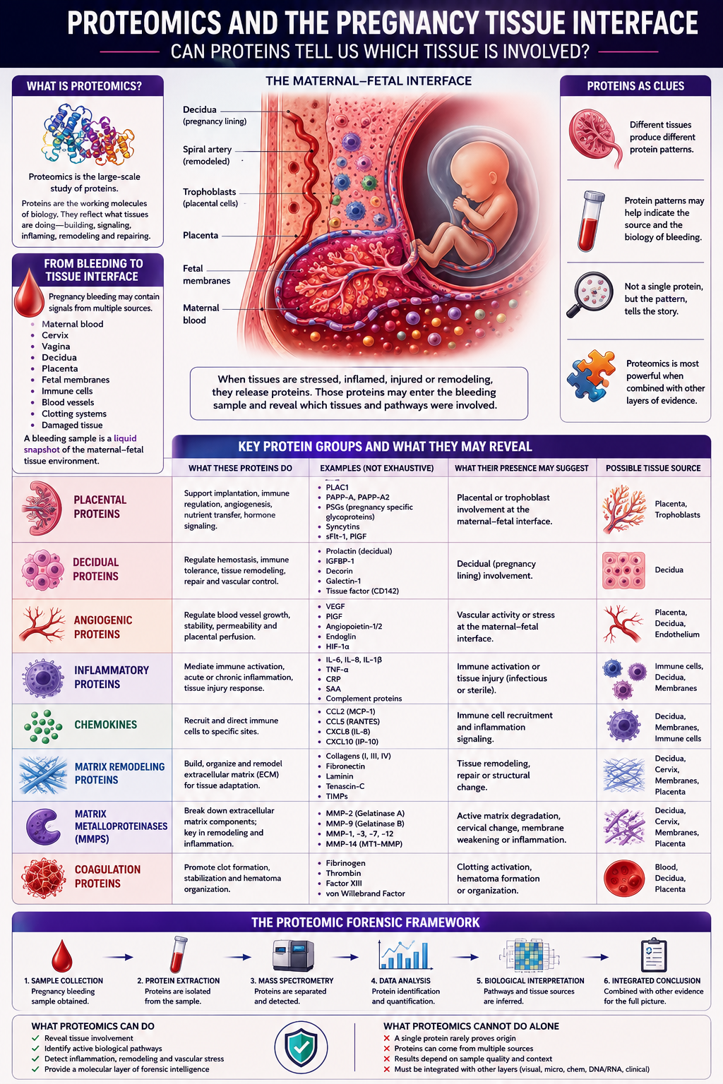
When these tissues are stressed, inflamed, injured or remodeling, they may release proteins.
Those proteins may enter the bleeding sample.
This means a sample of pregnancy bleeding may contain a protein fingerprint of the tissue interface where the event occurred.
The sample is not only blood.
It may be a liquid snapshot of the maternal-fetal tissue environment.
What Is Proteomics?
Proteomics is the study of many proteins at once.
Instead of measuring one marker, proteomics asks:
Which proteins are present?
How abundant are they?
Which biological pathways do they represent?
Which tissues may have released them?
Which processes are activated?
This is important because pregnancy bleeding is unlikely to be explained by one protein.
It is more likely to involve patterns.
A pattern of inflammatory proteins may suggest immune activation.
A pattern of matrix-remodeling proteins may suggest tissue breakdown.
A pattern of placental proteins may suggest placental-interface involvement.
A pattern of angiogenic proteins may suggest vascular stress.
Proteomics is therefore not just a list of proteins.
It is a way to read biological activity.
Placental Proteins
The placenta is one of the most protein-active organs in pregnancy.
It releases proteins into maternal circulation and into the local maternal-fetal interface.
Some placental proteins are involved in:
- implantation
- angiogenesis
- immune regulation
- nutrient transport
- vascular remodeling
- inflammation
- hormone signaling
If placental proteins appear in a bleeding sample, they may suggest that placental or trophoblast-associated tissue contributed to the sample.
Relevant protein groups may include:
- placental growth factor-related proteins
- pregnancy-associated plasma proteins
- trophoblast-derived proteins
- angiogenic regulators
- placental extracellular vesicle proteins
- immune-modulating placental proteins
The key point is not that one placental protein proves the source.
The key point is that a placental protein pattern may suggest placental-interface involvement.
Decidual Proteins
The decidua is the pregnancy-modified uterine lining.
It is not passive.
It controls hemostasis, immune tolerance, implantation, tissue remodeling and communication with the placenta.
During bleeding, decidual tissue may contribute proteins involved in:
- clotting
- inflammation
- immune regulation
- extracellular matrix remodeling
- tissue repair
- vascular control
A decidual protein signature may suggest that the bleeding involved the pregnancy lining rather than only the cervix or vagina.
This is especially important for hematoma-like bleeding.
A decidual hematoma is not just a collection of old blood.
It may be surrounded by active decidual tissue responding to blood, injury, inflammation and repair.
Angiogenesis: Blood Vessel Signaling
Pregnancy depends on blood vessel remodeling.
The maternal-fetal interface is built around controlled vascular change.
Proteins involved in angiogenesis may therefore be highly relevant.
Angiogenesis-related proteins include signals that regulate:
- blood vessel growth
- vessel stability
- endothelial function
- placental perfusion
- vascular leakage
- tissue oxygenation
If a bleeding sample contains abnormal angiogenic patterns, it may suggest vascular stress or placental-interface dysfunction.
Important concepts include:
- pro-angiogenic signaling
- anti-angiogenic signaling
- endothelial activation
- vascular instability
- placental perfusion stress
This matters because bleeding may sometimes reflect local vascular fragility rather than a simple mechanical event.
Inflammatory Proteins
Inflammation is one of the central biological themes in pregnancy bleeding.
Proteomics can detect inflammatory proteins that may reflect:
- acute immune activation
- chronic inflammation
- infection-associated inflammation
- sterile inflammation
- tissue injury
- hematoma response
Inflammatory protein groups may include:
- cytokines
- chemokines
- complement proteins
- acute phase proteins
- immune-cell activation proteins
- tissue injury proteins
Inflammation does not always mean infection.
Blood itself can trigger inflammation when it leaves the circulation and enters tissue spaces.
Old blood, heme, iron and damaged cells may all activate inflammatory pathways.
This means a hematoma can become biologically active even if it is not infected.
Chemokines and Immune Recruitment
Chemokines are proteins that guide immune cells.
They act like biological traffic signals.
They help determine which immune cells enter a tissue and where they go.
A bleeding sample rich in chemokines may suggest that the tissue was actively recruiting immune cells.
This could fit with:
- decidual inflammation
- infection-associated inflammation
- hematoma cleanup
- membrane stress
- tissue repair
- cervical remodeling
Microscopy may show immune cells.
Proteomics may show why they were recruited.
This is a major advantage of protein analysis.
It does not merely show what is present.
It can reveal the signaling logic behind what is present.
Matrix Remodeling
Tissues are not static.
They are built from extracellular matrix — collagen, elastin, proteoglycans and other structural components.
For pregnancy to progress, tissues must remodel in controlled ways.
The cervix remodels.
The decidua remodels.
The placenta invades and adapts.
The fetal membranes stretch and maintain strength.
Blood vessels transform.
If remodeling becomes excessive, inflammatory or poorly regulated, tissue integrity may be affected.
A bleeding sample may contain proteins related to matrix remodeling, such as:
- collagen fragments
- extracellular matrix proteins
- tissue inhibitors of metalloproteinases
- matrix metalloproteinases
- adhesion proteins
- basement membrane fragments
These proteins may help indicate whether tissue breakdown or repair was part of the bleeding event.
Matrix Metalloproteinases
Matrix metalloproteinases, often called MMPs, are enzymes that break down extracellular matrix.
They are essential in normal biology.
But when overactive, they may contribute to tissue weakening.
MMPs are relevant to:
- cervical remodeling
- membrane weakening
- inflammation
- placental invasion
- tissue repair
- extracellular matrix degradation
In the context of pregnancy bleeding, MMP-related patterns could suggest active tissue remodeling at the maternal-fetal interface.
This is particularly interesting for pathways involving:
- fetal membrane weakening
- decidual inflammation
- cervical change
- preterm birth biology
MMPs are not automatically harmful.
But their pattern and context may matter.
Proteins as Tissue Clues
Different tissues produce different protein patterns.
This raises the possibility of protein-based tissue-of-origin mapping.
A sample dominated by cervical proteins may suggest cervical involvement.
A sample containing decidual proteins may suggest pregnancy lining involvement.
A sample enriched with placental proteins may suggest placental-interface contribution.
A sample rich in membrane-remodeling proteins may suggest fetal membrane stress.
A sample rich in inflammatory proteins may suggest immune activation.
A sample rich in clotting and fibrin-related proteins may suggest coagulation and hematoma organization.
This is where proteomics becomes forensic.
It asks:
Which tissue left its protein signature in the sample?
Fresh Bleeding Versus Tissue-Interface Bleeding
Fresh bleeding from a superficial source may contain mostly blood proteins.
A more complex maternal-fetal interface bleed may contain:
- blood proteins
- decidual proteins
- placental proteins
- inflammatory proteins
- matrix-remodeling proteins
- coagulation proteins
- tissue injury proteins
This difference matters.
Both samples may look like vaginal bleeding.
But proteomically they may represent very different biological events.
That is the central value of this approach.
Proteins may reveal complexity that the eye cannot see.
The Proteomic Forensic Framework
A structured proteomics analysis could ask:
Blood Proteins
Is the sample mainly peripheral-type blood?
Placental Proteins
Is there evidence of placental or trophoblast contribution?
Decidual Proteins
Is the pregnancy lining involved?
Angiogenic Proteins
Is there vascular stress or abnormal vessel signaling?
Inflammatory Proteins
Is immune activation present?
Chemokines
Are immune cells being recruited?
Matrix Proteins
Is tissue structure being remodeled or degraded?
MMPs
Are matrix-degrading enzymes activated?
Coagulation Proteins
Is clot organization or hematoma biology present?
Together, these protein groups may help reconstruct the bleeding event.
Why Proteomics Is Different From Chemistry
Chemistry tells us about molecules and processes.
Proteomics tells us which protein systems are active.
That distinction matters.
pH, lactate, iron and oxidative stress markers describe the chemical environment.
Proteins can point more directly toward biological pathways and tissue involvement.
For example:
- angiogenic proteins may suggest vascular or placental biology
- MMPs may suggest matrix remodeling
- chemokines may suggest immune recruitment
- placental proteins may suggest trophoblast contribution
- decidual proteins may suggest pregnancy lining involvement
Proteomics is therefore one step closer to tissue interpretation.
What Proteomics Cannot Tell Us Alone
Proteomics is powerful, but it is not magic.
Proteins can come from multiple sources.
Many proteins are shared across tissues.
A single protein rarely proves origin.
Protein levels can be affected by:
- sample age
- dilution
- contamination
- clotting
- degradation
- collection method
- local tissue exposure
- inflammation
- infection
Therefore, proteomics should not be interpreted alone.
It should be integrated with:
- visual appearance
- microscopy
- microbiology
- chemistry
- DNA/RNA
- clinical context
The strength lies in pattern recognition.
The Central Question
The traditional clinical question is:
“How much bleeding is there?”
The proteomic question is:
“Which tissues and biological pathways left protein evidence in the sample?”
That is a different way of thinking.
Pregnancy bleeding becomes not only an event to observe, but a sample to decode.
Proteins may help reveal whether the event involved:
- blood only
- decidua
- placenta
- membranes
- inflammation
- vascular stress
- matrix breakdown
- clot organization
- tissue repair
Conclusion: The Fifth Layer of Evidence
Visual forensics asks:
What does the bleeding look like?
Microscopy asks:
What cells and tissues are present?
Microbiology asks:
What organisms and ecosystems are involved?
Chemistry asks:
What molecular environment exists in the sample?
Proteomics asks:
Which tissue pathways are active?
This is why proteomics may become one of the most important layers in Pregnancy Bleeding Intelligence.
Proteins are close to function.
They show not only what is present, but what the tissues may have been doing.
If pregnancy bleeding is a biological message, proteins may be some of its clearest words.
They may help us move from seeing blood to understanding the tissue interface behind it.
References and Resource
1. A Spatially Resolved Timeline of the Human Maternal–Fetal Interface Nature, July 2023.
This study used spatial proteomics and transcriptomics to map the human maternal–fetal interface during the first half of pregnancy, including decidua, trophoblasts, immune cells and vascular remodeling.
2. Proteome-Defined Changes in Cellular Pathways for Decidua and Trophoblast in Early Pregnancy Scientific Reports, February 2022.
This proteomics study compared decidual and trophoblast tissue from different early pregnancy outcomes, showing how protein patterns can reflect changes at the maternal–fetal interface.
3. Extracellular Matrix and Pregnancy: Functions and Opportunities Caught in the Net Reproductive Biology and Endocrinology, 2025.
A review of how extracellular matrix remodeling supports implantation, placentation, vascular adaptation and immune activity during pregnancy.
4. Insights Into the Immunomodulatory Regulation of Matrix Metalloproteinase at the Maternal–Fetal Interface During Early Pregnancy and Pregnancy-Related Diseases Frontiers in Immunology, January 2023.
A review focused on matrix metalloproteinases, immune regulation and tissue remodeling at the maternal–fetal interface, including their possible relevance as biomarkers in pregnancy disorders.
5. Placenta-Derived Angiogenic Proteins and Their Contribution to the Pathogenesis of Preeclampsia Angiogenesis, December 2014.
A review of placental angiogenic proteins, including factors involved in blood vessel growth, placental perfusion and vascular dysfunction in pregnancy complications.