

What color, texture, clots and visible fragments may reveal before laboratory testing
The first analysis of pregnancy bleeding does not begin in a laboratory.
It begins with the eye.
Before microscopy, microbiology, chemistry, proteomics or DNA analysis, there is a simple observation:
What does the bleeding look like?
In clinical care, visible bleeding is often described quickly: light, moderate, heavy, red, brown, with or without clots.
But from a forensic perspective, the visual appearance of bleeding may be treated as the first layer of information.
It may offer clues about timing, origin, tissue involvement and biological process.
This article focuses only on that first layer:
What can we learn from the visual appearance of vaginal bleeding during mid-pregnancy?
Not as a final diagnosis.
But as a structured way to observe.
Why Visual Forensics Matters
A bleeding sample is not just a volume of blood.
It is material that has come from somewhere, changed over time, passed through tissue and exited the body.
By the time it is seen, it may carry visible clues:
- whether it is fresh or old
- whether it is thin or coagulated
- whether mucus is present
- whether clots have formed
- whether tissue-like fragments are visible
- whether fluid has diluted it
- whether the pattern suggests active flow or delayed drainage
Visual inspection cannot answer everything.
But it can help ask better questions.
A forensic framework begins with disciplined observation.
The Four Visual Questions
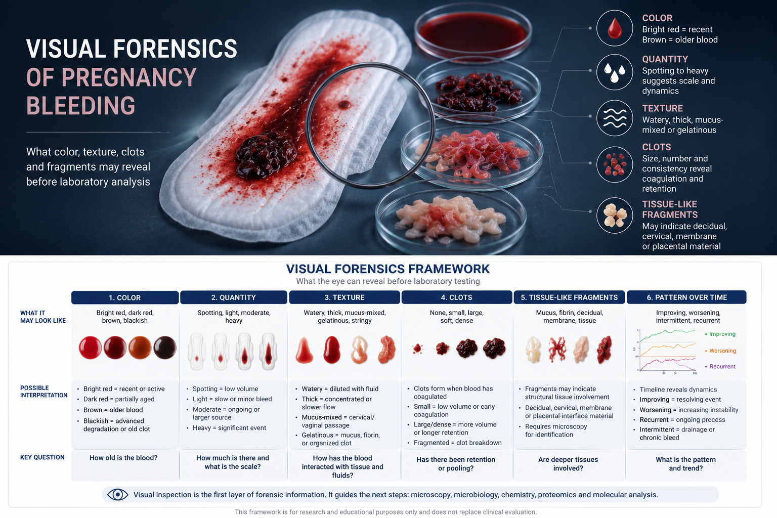
When looking at pregnancy bleeding, four basic questions matter:
- What color is it?
- How much is there?
- What is its texture or consistency?
- What is mixed into it?
These questions may sound simple.
But they are powerful because they create the first map of the bleeding event.
1. Color: The First Clue About Timing
Color is one of the most obvious visible features.
Bright Red Blood
Bright red blood usually suggests fresher bleeding.
It may indicate that blood has recently left a vessel or tissue surface and has not yet undergone much oxidation or degradation.
Possible visual interpretation:
- recent bleeding
- active bleeding
- faster movement out of the body
- less time retained in the reproductive tract
Bright red blood does not automatically mean danger, but it often suggests a more current event.
Dark Red Blood
Dark red blood may suggest blood that is older than bright red blood but not fully degraded.
It may have spent more time in contact with tissue, mucus or clot before exiting.
Possible visual interpretation:
- slower bleeding
- partial retention
- mixed fresh and older blood
- blood exposed to lower oxygen conditions
Brown Blood
Brown bleeding often suggests older blood.
The brown color usually reflects oxidation and breakdown of hemoglobin inside red blood cells.
Possible visual interpretation:
- previous bleeding event
- old blood slowly draining
- resolving hematoma
- retained blood now being expelled
- chronic or intermittent bleeding
In mid-pregnancy, brown bleeding may be especially relevant when considering hematoma-like processes, where blood may collect, remain in place, degrade and then later drain.
Blackish or Coffee-Ground Material
Very dark brown or blackish material may suggest more advanced blood degradation.
Possible visual interpretation:
- older retained blood
- organized clot
- hematoma drainage
- degraded red blood cells
- pigment-rich material
This does not determine the cause, but it strengthens the idea that the blood may not be from a brand-new event.
2. Quantity: The First Clue About Scale
Volume matters, but it should be described carefully.
Spotting
Small spots on tissue, underwear or a liner may suggest low-volume bleeding.
Possible visual interpretation:
- minor surface bleeding
- cervical irritation
- small draining hematoma
- old residual blood
- intermittent leakage
Light Bleeding
Light bleeding may be more than spotting but not enough to saturate pads quickly.
Possible visual interpretation:
- slow ongoing bleeding
- mild active bleeding
- drainage of older material
- cervical or decidual source
Moderate Bleeding
Moderate bleeding suggests a larger or more persistent event.
Possible visual interpretation:
- active bleeding source
- hematoma evacuation
- placental-interface involvement
- cervical change
- inflammatory or tissue-disruption process
Heavy Bleeding
Heavy bleeding visually suggests a significant event.
Possible visual interpretation:
- active vascular bleeding
- larger hematoma release
- placental-interface disruption
- clot passage
- need for urgent clinical evaluation
In a forensic framework, quantity is not just “how much.”
It is a clue about the scale and dynamics of the bleeding process.
3. Texture: Flowing, Watery, Thick or Gelatinous
Texture can reveal how the blood has behaved.
Thin or Watery Blood
Watery blood may suggest dilution.
Possible sources of dilution include vaginal secretions, cervical mucus or other fluid.
Possible visual interpretation:
- blood mixed with fluid
- lower concentration of red blood cells
- possible membrane/fluid question
- cervical or vaginal mixing
Thick Blood
Thick blood may suggest slower flow, clotting or concentration.
Possible visual interpretation:
- blood has had time to coagulate
- slower movement
- partial retention
- older bleeding material
Mucus-Mixed Blood
Mucus-mixed blood often appears stringy, slippery or gelatinous.
Possible visual interpretation:
- cervical passage
- cervical mucus contribution
- blood traveling through cervical canal
- local cervical irritation or change
Mucus does not prove that the cervix is the source, but it tells us the blood has interacted with cervical or vaginal secretions.
Gelatinous or Stringy Material
Gelatinous material may reflect mucus, fibrin or partially organized clot.
Possible visual interpretation:
- clot organization
- cervical mucus
- fibrin formation
- retained blood beginning to structure itself
4. Clots: Evidence of Coagulation and Retention
Clots are important visual evidence.
They tell us that blood has had time to coagulate.
Small Clots
Small clots may occur when bleeding is slow or intermittent.
Possible visual interpretation:
- low-volume coagulation
- minor retained blood
- mixed old and new bleeding
- early clot formation
Large Clots
Large clots suggest a greater volume of blood, longer retention or more substantial coagulation.
Possible visual interpretation:
- larger bleeding episode
- pooled blood
- hematoma evacuation
- significant retained material
Soft Clots
Soft, fresh-looking clots may suggest relatively recent coagulation.
Dense, Dark Clots
Dense dark clots may suggest older retained blood or more advanced organization.
Fragmented Clots
Fragmented clots may indicate that an older clot is breaking down and being expelled in pieces.
In forensic terms, clots are not just “clots.”
They are evidence of time, volume, coagulation and mechanical movement.
5. Tissue-Like Fragments
Visible fragments are especially important.
They may appear as pale, gray, pink, red-brown or stringy material mixed with blood.
Possible visual categories include:
- mucus fragments
- fibrin strands
- clot fragments
- decidual tissue
- cervical tissue
- membrane-like material
- placental-interface debris
- nonspecific cellular debris
Tissue-like material cannot be identified reliably by eye alone.
But its presence changes the question.
The sample is no longer only blood.
It may contain structural tissue information.
That means microscopy, histology or molecular analysis could become much more informative.
6. Mixed Old and Fresh Blood
One of the most informative visual patterns is mixed color.
For example:
- brown blood with bright red streaks
- dark clots with fresh red fluid
- old brown discharge followed by red bleeding
- red bleeding followed by brown spotting
This may suggest layered timing.
Possible interpretation:
- old bleeding is draining
- new bleeding has started
- a hematoma is intermittently emptying
- chronic bleeding with acute episodes
- clot breakdown with renewed flow
This is important because pregnancy bleeding may not be a single event.
It may be a sequence.
Visual appearance can sometimes show that sequence.
7. Pattern Over Time
A single observation is useful.
A timeline is better.
The pattern over hours or days may reveal dynamics.
Improving Pattern
Bright red → dark red → brown → stops
Possible interpretation:
- acute bleed resolving
- old blood clearing
- no obvious ongoing fresh bleeding
Worsening Pattern
Brown spotting → red bleeding → clots
Possible interpretation:
- old bleed followed by new active bleeding
- increasing instability
- hematoma expansion or release
- new tissue disruption
Intermittent Pattern
Brown bleeding on and off for days
Possible interpretation:
- slow drainage
- chronic retained blood
- small recurrent bleeds
- hematoma-like process
Recurrent Fresh Episodes
Repeated bright red bleeding
Possible interpretation:
- repeated active bleeding
- unstable tissue source
- cervical or placental-interface involvement
- ongoing vascular fragility
In forensic terms, time-series observation is more powerful than a single snapshot.
8. The Visual Matrix
A practical visual framework could look like this:
| Visual feature | Possible information |
|---|---|
| Bright red | More recent or active bleeding |
| Dark red | Partly aged blood |
| Brown | Older blood, degradation, retained blood |
| Blackish material | Advanced degradation or old clot |
| Watery | Dilution with fluid or secretions |
| Thick | Concentrated blood or clotting |
| Mucus-mixed | Cervical/vaginal passage or mucus contribution |
| Small clots | Coagulation, low-volume retention |
| Large clots | Larger volume, pooling, hematoma release |
| Tissue-like fragments | Possible structural tissue involvement |
| Mixed brown and red | Old bleeding plus newer bleeding |
| Recurrent pattern | Possible ongoing or repeated process |
This matrix does not diagnose.
It organizes observation.
9. What Visual Inspection Cannot Replace
Visual forensics is not a replacement for clinical assessment.
It cannot confirm:
- exact source
- fetal condition
- placental position
- cervical status
- infection
- membrane rupture
- genetic or molecular mechanisms
But that is not the point.
The point is that visual inspection is the first evidence layer.
It helps determine what questions should be asked next.
If brown blood suggests old bleeding, the next question becomes:
What made the old blood accumulate?
If clots are present, the next question becomes:
Where did coagulation occur?
If mucus is present, the next question becomes:
Did the bleeding pass through or originate near the cervix?
If tissue fragments are present, the next question becomes:
What tissue is this?
A good forensic framework does not jump to conclusions.
It generates better hypotheses.
10. Why This Matters for Research
In research, visual information is often underused.
Samples are processed, centrifuged, frozen or discarded without preserving the original visual context.
But the visual phenotype may matter.
A brown, thick, clot-rich sample is not the same as a thin, bright red sample.
A mucus-rich sample is not the same as a tissue-fragment-rich sample.
A single “pregnancy bleeding sample” category may hide many different biological events.
Future research should classify pregnancy bleeding samples by visual phenotype before deeper analysis.
Possible visual categories:
- fresh red bleeding
- old brown bleeding
- mixed fresh and old bleeding
- mucus-rich bleeding
- clot-rich bleeding
- tissue-fragment bleeding
- watery blood
- recurrent low-volume bleeding
- single high-volume bleeding
These categories could then be compared with microscopy, microbiology, chemistry, proteomics, DNA/RNA and clinical outcomes.
That is how visual forensics becomes scientifically useful.
11. The Link to Hematoma
Visual forensics is especially relevant for pregnancy hematoma.
A hematoma is not just bleeding.
It is blood that has collected somewhere.
Once blood collects, it can age, clot, organize, inflame, degrade and eventually drain.
This means hematoma-associated bleeding may appear:
- dark
- brown
- clot-rich
- intermittent
- mixed with degraded material
- sometimes followed by fresh red bleeding
The visual pattern may help distinguish a simple fresh bleed from old stored blood being released.
That distinction is central to understanding hematoma biology.
12. From Visual Signal to Biological Question
The most important shift is conceptual.
Instead of asking only:
“How much bleeding is there?”
we should also ask:
“What does the bleeding look like?”
And then:
“What does that appearance suggest biologically?”
Visual observation can point toward:
- timing
- retention
- coagulation
- degradation
- mucus involvement
- tissue involvement
- fluid mixing
- repeated events
This is the first step in moving from symptom description to biological interpretation.
Conclusion: The First Layer of Evidence
Visual forensics is the simplest layer of pregnancy bleeding analysis.
It requires no advanced technology.
Only structured observation.
Color, quantity, texture, clots, mucus and fragments may all carry information.
They do not provide a final diagnosis.
But they can help reconstruct the event.
A bleeding sample has a history.
It came from somewhere.
It changed over time.
It mixed with tissue and fluid.
It may have clotted, degraded or carried fragments with it.
The visual appearance is the first trace of that history.
Before we ask what the microscope, chemistry or DNA can reveal, we should ask what the eye can already see.
Because in pregnancy bleeding, the first forensic instrument is observation.
References and Resource
- Bleeding During Pregnancy ACOG
Patient-friendly overview of pregnancy bleeding, including why bleeding should be reported and what broad causes clinicians consider - Antepartum Haemorrhage Green-top Guideline No. 63 Royal College of Obstetricians and Gynaecologists
Clinical guideline on bleeding after 24 weeks, including placenta previa, abruption, local cervical/vaginal causes, and unexplained bleeding - Vaginal Bleeding During Late Pregnancy MSD Manual Professional
Concise clinical reference covering evaluation of bleeding later in pregnancy and serious causes such as placental abruption, placenta previa and vasa previa - Placenta Previa MSD Manual Professional
Useful background on placenta previa as a key cause of second- and third-trimester bleeding, including why bleeding after 20 weeks requires careful assessment - Estimating the Age of Blood Stains and Its Application in Forensic Science Springer
Forensic reference on blood-age estimation, including why color and degradation can provide clues but are affected by environment and sample conditions